


Dermatology Clinic, University of Catania, AOU Policlinico-Vittorio Emanuele, Via Santa Sofia, 78, IT-95123, Catania, Italy. *E-mail: cldermct@gmail.com
Accepted Mar 27, 2017; Epub ahead of print Mar 30, 2017
Acne conglobata is a severe variant of inflammatory acne presenting with abscesses, cystic nodules, and sinus tracts. Comedones appearing in the late stage of the disease have been reported and defined as polyporus/fistulated comedones or secondary comedones (1, 2). We report here a patient with a large number of blackheads on his back. The patient was evaluated with dermoscopy and histopathology. The results were compared with those from our recent study of double-ended pseudocomedones in patients with hidradenitis suppurativa (HS) (3).
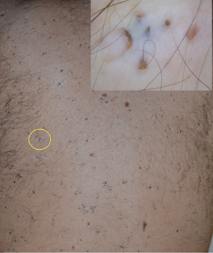
A 50-year-old man with a past history of acne conglobata presented clinically with more than 100 comedones spread diffusely on his back, mostly distributed in clusters, and arising on a whitish/fibrotic background (Fig. 1). No inflammatory lesions were seen. Ten clusters were selected randomly for polarized light ×10 dermoscopy (Dermlite Hybrid®, 3 Gen, San Juan Capistrano, CA, USA), all showing the same pattern: the presence of roundish, irregular depressions with enlarged openings centred by superficially located or deep-seated keratin debris and always arising on a whitish scar tissue background of variable depth and morphology (Fig. 1 insert). Histopathology of one lesion showed a horn-filled horizontally-oriented cavity lined with normal multilayered epithelium and surrounded by fibrosis. There was no evidence of hair follicle remnants.
Fig. 1. Hundreds of clustered comedones on the back in a patient with a previous history of acne conglobata. Insert: dermoscopy of a cluster (yellow circle) showing multiple roundish, irregular depressions with enlarged openings, centred by superficially located or deep-seated keratin debris and arising on a whitish scar tissue background (×10). The same pattern was observed in all clusters examined (n = 10).
The comedones in this case were different from the primary comedones of acne, which represent the first step of a later development into inflammatory lesions. Primary comedones usually appear as single/multiple, follicular-based black-headed papules. In our patient, in addition to the different clinical presentation, i.e. clustering of multiple enlarged blackheads within areas of cicatricial tissue, all comedones were preceded by severe acne, and histopathology of one lesion showed no evidence of hair follicle.
Based on these observations, these lesions should be considered secondary comedones, representing the final, scarring outcome of severe acne, commonly acne conglobata, where they are generally observed on the back as hundreds of lesions. The literature in this field is scant and, according to the original description, each comedo is a member of a complex system of interconnected horn-filled galleries resulting from rupture, abscess formation and re-encapsulation of adjacent follicular units (2). This definition and pathogenetic interpretation of such lesions fit well with our findings.
We compared our results with those of 20 patients with HS showing numerous so-called double-ended pseudocomedones located in HS typical body areas (3). Interestingly, we found striking similarities (Fig. 2): both secondary comedones in acne conglobata and pseudocomedones in HS present as single/multiple lesions, often arranged in clusters, with negligible or absent inflammation and always surrounded by cicatricial tissue.
Fig. 2. Pseudocomedones on the pubic area in a woman affected by hidradenitis suppurativa. Insert: dermoscopy showing irregular openings of variable size and superficial and deep-seated keratin accumulations embedded in a whitish cicatricial tissue. The same pattern was observed in all lesions examined (n = 64) out of 20 patients.
Also, they share similar histopathological features, consisting of horn-filled cavities lined with multilayered epithelium with no evidence of follicle structures. This resemblance probably is not a coincidence. It is well-known that acne conglobata and HS may coexist (4), probably sharing some pathogenetic mechanisms, namely follicular occlusion, inflammation, and rupture followed by cicatricial rearrangement in the late stages.
In conclusion, secondary comedones, pseudocomedones, or, as suggested in a recent HS nomenclature review, “keratin-filled interconnected multipores” (5), should be viewed as the final outcome of repeated healing from relapsing inflammatory and destructive processes of the follicular unit, as observed in acne conglobata and HS.
The authors declare no conflicts of interest.


 Click to show fullsize
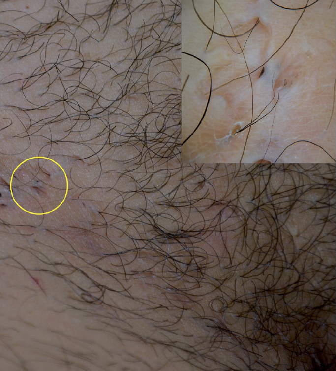 Click to show fullsize