


1Department of Dermatology, Venereology and Allergology, HELIOS St Elisabeth Hospital Oberhausen, University Witten/Herdecke, Josefstr. 3, DE-46045 Oberhausen, 2Institute of Virology, National Reference Center for Papilloma- and Polyomaviruses, University of Cologne, and 3Institute of Pathology, Mülheim an der Ruhr, Germany. E-mail: alexander.kreuter@helios-kliniken.de
#These authors contributed equally to this work.
Accepted Feb 20, 2018; Epub ahead of print Feb 28, 2018
Infections with human papillomaviruses (HPV) induce a heterogeneous spectrum of cutaneous and mucomembraneous lesions. Epithelial lesions caused by HPV-types of the genus alpha (e.g. anogenital warts/condylomata acuminata, intraepithelial neoplasias and invasive cancers) predominantly occur in the anogenital region. Depending on their oncogenic potential, alpha-HPVs can be divided into high-risk (e.g. HPV16 or HPV18) and low-risk types (e.g. HPV6 or HPV11). In contrast to high-risk HPVs, low-risk HPVs are only rarely capable of inducing high-grade dysplasias or anogenital cancers (< 1–10%) (1–3). We report here an immunocompetent patient with simultaneous development of benign condylomata acuminata and high-grade anal dysplasia induced by the same low-risk HPV-type, HPV42.
A 59-year-old woman was referred to our department because of a monofocal high-grade anal intraepithelial neoplasia (AIN grade 3; AIN3) that had been surgically removed during colonoscopy for routine colon cancer screening. The patient had no clinical and laboratory signs of immunodeficiency (HIV testing was negative and lymphocyte subpopulations including CD3+ T-cells, CD4+ T-cells, CD8+ T-cells, CD19+ B-cells, and natural killer (NK) cells were within normal ranges), no previous or current immunosuppressive medication, and no history of cervical or other HPV-related disease. At first clinical examination with high-resolution anoscopy (HRA) in our department, a new lesion suspicious for HPV-related disease was surgically removed, and was again histopathologically diagnosed as AIN3 (located at 7 o’clock in lithotomy position) (Fig. 1). At 2 further HRA-examinations 1 and 3 months thereafter, 2 more intra-anal lesions were surgically removed and histopathology revealed benign condyloma (at 9 o’clock) and AIN3 (at 3 o’clock), respectively. All 3 lesions had identical clinical HRA-features usually seen in benign intra-anal condyloma (dome-shaped papules with homogenous terminal capillaries and papillary structures), as reported previously (4). Characteristic HRA-signs of high-grade dysplasia, such as punctation, mosaicism, or neovascularization, were missing.
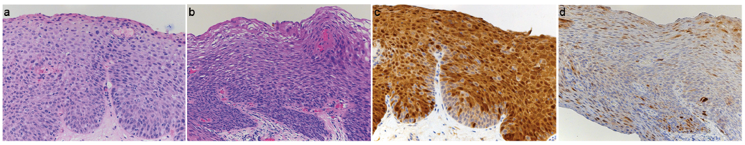
Fig. 1. Routine histopathology and p16INK4a-immunohistochemistry of the condyloma and of the first AIN3 lesion. (a) Atypical cells are present within the entire epithelium of the AIN3 lesion (haematoxylin and eosin (H&E) staining). (b) Classic histological features of condyloma, including hyperkeratosis, parakeratosis, and koilocytosis, are present (H&E staining) (c) “Band-like” p16INK4-positivity with both nuclear and cytoplasmatic staining within the entire lesion is present, characteristic for HPV-induced high-grade dysplasia (p16INK4a-immunostaining). (d) Negative p16INK4a-staining of the anal condyloma (focal p16INK4a-staining is considered as negative). Original magnification ×100.
HPV detection and typing from these 3 lesions was performed with 3 different alpha-HPV group-specific PCRs, respectively, followed by bead-based or reverse line blot hybridization, as described previously (4). Only low-risk type HPV42 was found in the 3 biopsies, and none of the other 38 low-risk- and high-risk alpha HPV-types covered by the 3 assays were detected. HPV42 DNA load was determined by real-time PCR with type-specific primers (fw: TGATACTGAAAATGCGCCTACAT; rev: CATAGAAACATTTTCCCTATTGTCTG) and locked nucleic acid probe no. 81 (GGCCCTGG; Cat. no.: 04689046001, Roche, Mannheim, Germany) using a Light Cycler 480 (Roche) (5). HPV42 load was expressed as HPV42 DNA copies per betaglobin-gene copy. Viral load determination showed HPV42 DNA loads well above 1 in all 3 samples: 64.7 HPV42 DNA copies per betaglobin gene copy in the first AIN3 lesion of January 2017 (7 o’clock in lithotomy position), 18.7 in the condyloma without dysplasia of February 2017 (9 o’clock), and 2.0 in the AIN3 lesion of April 2017 (3 o’clock). P16INK4a-immunohistochemistry was performed using the CINtec histology kit (Roche, Mannheim, Germany) according to the manufacturer’s instructions, and p16INK4a-staining, an indirect marker of HPV E7 oncogene expression, was evaluated as previously described (6). Strong p16INK4a-positivity was found in both AIN3 lesions, whereas p16INK4a-staining was negative in the anal condyloma (focal p16INK4a-staining is considered as negative) (Fig. 1).
Next, in situ hybridization was performed according to previously reported protocols (7). The hybridization probe was generated with the DIG-Nick-Translation Mix (Roche) using pSP64-HPV42 as template and pSP64 as negative control (8). Slides were developed with the TSA Plus Fluorescein Kit (PerkinElmer, Waltham, MA, USA). In accordance with the high HPV42 DNA loads measured by real-time PCR, in situ hybridization confirmed high HPV42 DNA levels, both in the AIN3 lesions and in the condyloma (Fig. S1).
To the best of our knowledge, this is the first investigation demonstrating that a low-risk HPV-type, in our case HPV42, may simultaneously induce benign condyloma and high-grade anal dysplasia in the same patient within a short period of time. Importantly, both types of lesions developed within a few weeks; an observation that is in line with a previous observation of our group on anal cancer and its precursors induced by high-risk HPV-types (9). The induction of anogenital carcinomas by low-risk HPVs is very rare. Among 13,328 HPV-induced carcinomas evaluated in a global study, only 57 were associated with a single low-risk HPV infection and only 4 (3 cervical cancers and 1 anal cancer) were induced by HPV42 (1). Similarly, single low-risk HPV-type infections were found in less than 1% of 1,739 squamous cell cervical cancers from 9 countries and HPV42 was not detected (2). In a study of anal carcinomas worldwide, a HPV42 single infection has been found in one of 438 (0.2%) cases (10). HPV42 was originally isolated from a benign vulvar papilloma and has been found in 1–2% of condylomas in recent studies (11–13). We and others have recently detected HPV42 mono-infections in AIN-containing intra-anal condylomas of HIV-positive patients (4, 14). In contrast to high-risk HPVs, the E6 oncoprotein of HPV42 does not degrade the tumour suppressor p53, but its E7 oncoprotein seems to be able to interact with the tumour suppressor protein pRB, as shown by strong p16INK4a-expression in HPV42-positive dysplasias and cancers (1). This is in contrast to anogenital malignancies (e.g. Buschke-Löwenstein tumors) induced by HPV6 or HPV11, that lack p16INK4a-expression and histologically show features of verruco-papillary tumours (1).
Interestingly, the 3 anal lesions in our patient were clinically indistinguishable and showed typical HRA-features of benign condylomas. This is in line with previous findings of our group and reports of others, showing that lesions with clinical characteristics of condylomas can contain anogenital dysplasia and that these dysplasias can be induced by low-risk HPV-types, both in HIV-positive men who have sex with men (4, 14) and HIV-negative men and women (15).
In summary, we demonstrate that a low-risk genus alpha HPV-type, HPV42, can induce both benign condyloma and anal high-grade dysplasia, a cancer precursor, in the same individual within a short period of time. It should be noted that clinically benign-appearing lesions caused by low-risk HPV-types may harbour severe dysplasia, not only in immunosuppressed patients (4), but also occasionally in immunocompetent individuals.
The authors thank Monika Junk and Nabila Ristow for excellent technical assistance. The HPV42 clone (pSP64-HPV42) was originally isolated by Michel Favre at the Institute Pasteur, Paris, France and was obtained from the International HPV Reference Center, Karolinska Institute, Stockholm, Sweden.
Funding sources: HPV testing was funded by the German National Reference Center for Papilloma- and Polyomaviruses (German Federal Ministry of Health, grant number 1369-401). MH was supported by the German Cancer Aid (Deutsche Krebshilfe, grant number 111087).
The authors have no conflicts of interest to declare.


 Click to show fullsize