


1Department of Dermatology, and 2Department of Geriatrics, The First Hospital of China Medical University, 155 North Nanjing Street Shenyang 110001, China. *E-mails: hongduochen@hotmail.com; chundihe@163.com
Accepted Apr 2, 2019; E-published Apr 2, 2019
Tylosis with esophageal cancer (TOC) is an extremely rare autosomal dominant disorder characterized by early-onset palmar and plantar hyperkeratosis, oral leukoplakia, and a high risk of esophageal cancer (1, 2). The mutation of inactive rhomboid protease RHBDF2 gene has been reported as the underlying cause of TOC (3). Up till now, there are only 3 studies on RHBDF2 gene mutations in TOC reported (3–5). Here for the first time, we identified a novel c.589C>A mutation of RHBDF2 gene from a Chinese family with TOC by whole exome sequencing. We found that the mutation in the RHBDF2 gene altered RHBDF2 protein localization in tylotic skin. We also found that both sporadic and tylotic esophageal cancer types present different expression of the RHBDF2 compared to the normal controls.
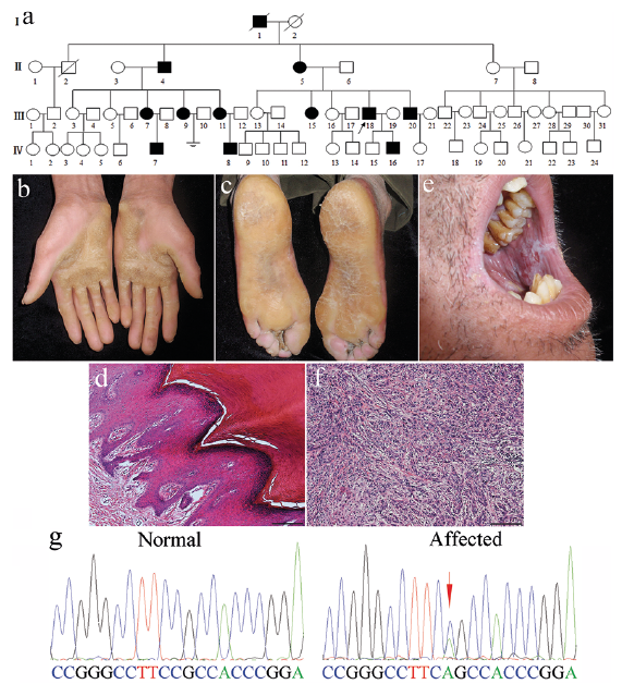
The Chinese family of interest has 65 individuals spanning 4 generations, of which 12 were affected (Fig. 1a). The proband (III18) is a 51-year-old man who has been affected by palmar (Fig. 1b) and plantar (Fig. 1c, d) hyperkeratosis since he was 9 years. He was diagnosed with oral leukoplakia (Fig. 1e) and esophageal cancer (Fig. f) at the age of 48. Other 7 family members (I1, II4, II5, III7, III9, III11 and III18) affected by the palmar and plantar hyperkeratosis have also been diagnosed with esophageal cancer. Four family members (III20, IV7, IV8 and IV16) were affected by the palmar and plantar hyperkeratosis, without esophageal cancer diagnosis so far. For complete details see Appendix S1.
Written informed consent was obtained from all subjects and the Institutional Review Board and the Ethics Committee of The First Hospital of China Medical University provided approval for this study. Peripheral blood samples were collected from the TOC (tylosis with esophageal cancer) family members and 100 unrelated healthy individuals of Chinese origin as controls. Three affected (III11, III18 and IV16) and one unaffected individuals (III5) were subjected to the whole exome sequencing. Genomic DNAs were isolated and randomly fragmented. Whole exome sequencing was performed on all samples using Agilent SureSelect All Exon V6 kit and paired-end 150 bp sequencing on the Illumina NovaSeq platform. Sequencing reads were aligned to the GRCh37 reference genome using Burrows Wheeler Aligner (BWA). Reads that aligned to exon regions were collected for mutation identification and subsequent analysis. SAMtools mpileup and BCFtools were used to do variant calling and identify SNP and indels. The data that mapped to the targeted region chr17q25 had a mean depth of 144.01-fold, and 99.95% of the targeted bases were covered. Finally, 4 variants in 3 genes, including RHBDF2 gene: c.502C>A and c.589C>A; ITGB4 gene: c.2207G>T; and CDK3 gene: c.803A>G, with the greatest likelihood of relevance were checked by Sanger sequencing. Only the heterozygous mutation c.589C>A in exon 6 of the RHBDF2 gene co-segregated with the phenotype and was observed neither in unaffected family members nor in 100 unrelated Chinese controls (Fig. 1g).
Using DNAMAN software (Lynnon BioSoft, Canada), we confirmed that mutation c.589C>A lead to the amino acid alteration (p.Arg197Ser). Plantar skin and the whole layer of esophageal mucosa samples were collected from 3 affected family members (III9, III11 and III18), and 3 unaffected family members (III10, III12 and III19). Biopsies of the whole layer of the mucosa were also obtained from 3 sporadic squamous esophageal cancers patients. We found that RHBDF2 appeared to localize predominantly on the cell membrane in sections from normal skin, whereas the localization was mostly cytoplasmic in skin sections from patients with tylosis (Fig. S1a). Immunohistochemical staining potentially indicated that the expression of RHBDF2 in both sporadic and tylotic esophageal cancer types is strongly cytoplasmic compared to the normal esophageal mucosa (Fig. S1b). These data are consistent with previous reports (3).
Fig. 1. (a) Family pedigree. (b) Clinical images of the proband’s palmar, (c) plantar and (e) oral leukokeratosis. (d) Skin biopsy from the proband’s plantar, and (f) mucosa biopsy from the proband’s esophageal cancer tissue. Scale bars represent 100 μm. (g) c.589C>A mutation in the exon 6 of RHBDF2 gene from normal controls and affected members of the family.
Previous studies have localized the TOC locus to a small genomic interval within the chromosomal region chr17q25.1-q25.2 and excluded several candidate genes (6). Recently, it was reported that after extending and revising the limits of the TOC minimal region, two heterozygous missense mutations in exon 6 of the RHBDF2 gene were found to segregate with the TOC syndrome: a c.557T>C mutation in a UK family and a US family; a c.566C>T mutation in a German family (3). A c.562G>A mutation in a Finnish family of TOC and a c.562G>T mutation in an African family of TOC were also reported (4, 5).
RHBDF2 is a highly conserved, catalytically inactive protein belonging to the rhomboid family of intramembrane serine proteases. RHBDF2 has been linked to the regulation of epidermal growth factor (EGF) signaling (7). It was reported that the distribution of RHBDF2 in tylotic skin was different from that in normal skin, and immortalized tylotic keratinocytes had lower levels of total epidermal growth factor receptors (EGFR) and displayed a higher proliferative and migratory potential relative to normal cells, even when normal cells were stimulated with exogenous epidermal growth factor (3, 8). It would thus appear that EGFR signaling is dysregulated in tylotic cells. It was also demonstrated that TOC-associated mutations in RHBDF2 cause an increase in the maturation and activity of the multi-substrate ectodomain sheddase enzyme ADAM17 in epidermal keratinocytes, derived from TOC patients, resulting in significantly upregulated shedding of ADAM17 substrates, including EGF-family growth factors and pro-inflammatory cytokines. This activity is accompanied by an increased EGFR activity (7). Furthermore, it indicates an altered localization of RHBDF2 in both tylotic and sporadic squamous cell esophageal tumors (3). Substantial evidence suggests that RHBDF2 regulates the EGFR signaling path-way and its downstream signaling events, including development of tylosis and epithelial tumorigenesis, through an enhanced secretion of the EGFR ligand amphiregulin (AREG) (9, 10, 11). It indicates a tissue-specific role of the RHBDF2-AREG-ADAM17-EGFR pathway in TOC, using mouse models (9, 11).
Our study reports a novel mutation of the RHBDF2 gene from a large Chinese family with TOC with different RHBDF2 protein expression in tylotic skin, both sporadic and tylotic esophageal cancer types compared to corresponding normal controls. It suggests that the c.589C>A mutation in exon 6 of the RHBDF2 gene could lead to TOC development, which provides a new mutation to the RHBDF2 gene mutation database. The finding will, hopefully, also benefit research on sporadic esophageal cancers, in which loss-of-heterozygosity and deletions in the TOC locus have been detected (12).
We thank members of the family who participated in this project. This work was supported by grants from National Natural Science Fund of China (81602741), Science & Technology Fund of Liaoning Province (201501013) and Science & Technology Fund of Shenyang (17-230-9-15).
The authors have no conflicts of interest to declare.


 Click to show fullsize