


Department of Dermatology and Venereology, Copenhagen University Hospital, Bispebjerg Hospital, DK-2400 Copenhagen, Denmark. *E-mail: hellekl@dadlnet.dk
Accepted Jan 28, 2020; Epub ahead of print Jan 31, 2020
Acta Derm Venereol 2020; 100: adv00072
Syphilis is a systemic human infection caused by the spirochete Treponema pallidum (1). T. pallidum is an obligate human pathogen, which is renowned for its invasiveness and immune-evasiveness. Syphilis is divided into stages, based on the clinical findings and corresponding serology (2). Due to its many atypical clinical presentations, syphilis is known as “the great imitator”. However, textbooks often describe the primary lesion as solitary, painless and indurated. This description may mislead inexperienced physicians when syphilis presents atypically. From being almost eradicated, in recent years syphilis has re-emerged in Western countries, mainly among men who have sex with men (MSM) (1). Due to the increase in numbers of infected individuals, it is expected that doctors also will observe an increase in atypical presentations, such as the herpetiform manifestation of primary syphilis presented in this case series.
This case series is a small retrospective pilot sample of sporadically collected cases of patients diagnosed with herpetiform penile lesions of syphilis, in the period September 2008 to May 2017, at the Sexual Transmitted Infections (STI) Clinic, at Bispebjerg Hospital (BBH), Copenhagen, Denmark.
The study was approved by the Danish Data Protection Agency, the Capital Region of Denmark (reference number VD-2019-236).
At initial visit all patients are offered standard tests and examination for HIV, syphilis (blood sample), chlamydia and gonorrhoea. Hepatitis B and C screening is offered to high-risk groups, including MSM.
All T. pallidum PCR (TP PCR) results in this case report series were analysed at Statens Serum Institute, Copenhagen (SSI). Until January 2013, syphilis serology in Denmark was exclusively analysed at SSI using the following tests: Wasserman reaction (WR), rapid plasma reagin test (RPR), antiflagel IgG (AF-G) and antiflagel IgM (AF-M) and fluorescent treponemal antibody-absorption test (FTA-ABS). After January 2013, all blood samples for syphilis screening taken at BBH were sent to the Department of Clinical Biochemistry at BBH, and screened with Architect Syphilis TP assay (Abbott, Abbott Park, Chicago, Illinois, USA). In case of a positive screening test, the sample was sent for further analyses at the Department of Microbiology at Hvidovre Hospital, with RPR, T. pallidum particle agglutination assay (Serodia-TPPA, Fujirebio, Japan) and INNO-LIA Syphilis Score, Fujirebio, Europé, Belgium (line immunoassay).
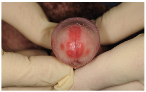
This case series includes 12 men, who presented with herpetiform penile ulcers, which were subsequently diagnosed as primary syphilis. In only one of these cases was the diagnosis of primary syphilis made at the first visit. This person had correspondingly 2 years prior to the actual infection been diagnosed with the exact same type of herpetiform penile lesions (Fig. 1). The time from appearance of the penile ulcers at the first visit to the clinic was between 1 day and 3 weeks.
Fig. 1. Herpetiform erosions in a 46-year-old HIV-negative homosexual man, Treponema pallidum PCR positive and herpes simplex virus PCR negative.
Eight of the 12 men (67%) were exclusively MSM with additionally 2/12 (16.7%) being bisexual. Two of the 12 men (16.7%) were HIV-infected, of whom one was MSM and one bisexual. The median age of the men was 41.2 years (range 26–64 years). Only 2/12 (16.7%) of the men had previously had syphilis.
All 12 men were initially tested with HSV PCR; only one tested positive, and was thus found to be co-infected with T. pallidum and herpes simplex virus type 2 (Fig. 2).
Fig. 2. Herpetiform erosions in a 36-year-old HIV-positive bisexual man, Treponema pallidum and herpes simplex virus PCR positive.
Based on the clinical presentation, HSV infection was the tentative diagnosis in 9/12 men, with descriptions such as “herpetiform erosions”, “herpetiform ulcers” or “suspicion of herpes genitalis”, whereas the remaining lesions were described as “flat ulcers” or “erosions”. Furthermore, 5 cases (42%) started treatment with aciclovir at the initial visit before the results of the syphilis serology or TP PCR were available. For 3/9 patients no tentative diagnosis was presented in the patient file at first visit. In 10 out of 12 men TP PCR was performed at the first visit, of which information was available in 9 of the 10 patient files, all being positive. All 12 men were treated with a single dose of benzathine penicillin G, 2.4 million units intramuscular (IM).
The presentation of all 12 patients in this case series differs from the typical solitary chancre often described in textbooks, by having 2 or more penile ulcers without induration. Among 9/12 men the tentative diagnosis on the day of presentation was herpes genitalis due to their clinical presentation. Similar findings have been documented in an Australian study (3). Of 183 men with a positive TP PCR, multiple anogenital ulcers where observed in 37.7%, and 49.2% having painful/tender ulcers, with no significant difference according to HIV status, only 2.7% had a concurrent positive HSV PCR (3). Previous studies have, in contrast, stated that multiple chancres is a more common presentation in patients with a coexisting HIV infection (4, 5). However, only 2/12 patients in the present case series were HIV-infected, This finding suggests that the herpetiform manifestation of primary syphilis with multiple ulcers, without concurrent HSV infection, is not a presentation observed more frequently among HIV-infected men. In addition, in a French study of 278 cases of genital ulcers, in which primary syphilis was the most common aetiology among 40% of the men, and in particular among 71% of MSM, no difference in the mean and median number of lesions or the size of ulcers was found according to HIV status (6). In a recent Spanish study, in which 31.1% of patients with primary syphilis had more than one ulcer, no association could be found with certain strain types or with HIV status (7)
In the secondary stage of syphilis mucosal lesions, such as condylomata lata or eroded syphilitic papules, may be observed (1). Based on the paraclinical findings in this report, we were able to distinguish the lesions found as primary syphilitic lesions. All men in this case series had either a positive TP PCR and subsequently seroconversion, or had low titres in non-treponemal tests, which subsequently increased (data not shown). Based on their clinical appearance and serological results all patients were diagnosed as being in the primary stage of syphilis.
This case report series has some limitations. First, the study of the included cases is retrospective and cases were selected sporadically. Secondly, the number of cases is small, and the findings should be reproduced in a larger study.
Previous studies have shown that, even among experienced physicians, the accuracy of clinical diagnosis of genital ulcer diseases, including primary syphilis, especially in men, is low compared with paraclinical analysis (8, 9).
All cases were diagnosed at the STI clinic at Bispebjerg Hospital, despite initial suspicion of an HSV infection. This could be explained by routine screening for STIs, including syphilis serology and clinical and serological follow-up in cases of genital ulcers. In less experienced settings, a risk of a delayed diagnosis, and thus progression to later stages of syphilis, would be of concern.
In conclusion, the present case series underlines the importance of creating awareness and continuous education of healthcare providers regarding atypical presentations of primary syphilis. This includes considering syphilis as a tentative diagnosis in patients presenting clinically with multiple herpetiform genital lesions to ensure rapid diagnosis and correct treatment of syphilis, thereby preventing progression to later stages and spread of the infection.


 Click to show fullsize
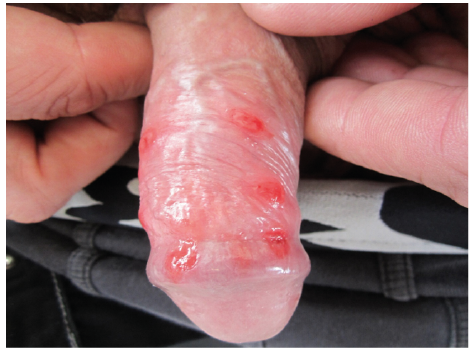 Click to show fullsize