


1Department of Dermatology, Monash University, Eastern Health, Box Hill, Victoria, 2Murdoch Childrens Research Institute, Melbourne, Victoria, 3School of Medicine, Griffith University, Gold Coast and Bond University Medical School, Robina, Queensland, 4Department of Paediatrics, The Royal Children’s Hospital, Melbourne, Victoria, 5Department of Paediatrics, University of Melbourne, Melbourne, Victoria, 6Queensland Institute of Dermatology and Veracity Clinical Research, Brisbane, Queensland, 7The Skin and Cancer Foundation Inc., Melbourne and Department of Dermatology, Alfred Health, Melbourne, Victoria, Australia, 8The School of Materials, The University of Manchester, Manchester, United Kingdom, and 9Department of Medicine, Campbelltown Hospital and School of Medicine, Western Sydney University, New South Wales, Australia
Although wool is commonly believed to cause irritant (non-immune) and hypersensitivity (immune) cutaneous reactions, the evidence basis for this belief and its validity for modern garments have not been critically examined. Publications from the last 100 years, using MEDLINE and Google Scholar, were analysed for evidence that wool causes cutaneous reactions, both immune-mediated (atopic dermatitis exacerbation, contact urticaria, allergic contact dermatitis) and non-immune-mediated (irritant contact dermatitis, itch). Secondary aims of this paper were to examine evidence that lanolin and textile-processing additives (formaldehyde, chromium) cause cutaneous reactions in the context of modern wool-processing techniques. Current evidence does not suggest that wool-fibre is a cutaneous allergen. Furthermore, contact allergy from lanolin, chromium and formaldehyde is highly unlikely with modern wool garments. Cutaneous irritation from wool relates to high fibre diameters (≥ 30–32 µm). Superfine and ultrafine Merino wool do not activate sufficient c-fibres to cause itch, are well tolerated and may benefit eczema management.
Key words: wool; allergy; atopic dermatitis; contact dermatitis; irritant dermatitis.
Accepted Mar 22, 2017; Epub ahead of print Mar 28, 2017
Acta Derm Venereol 2017; 97:
Corr: Dr Michaela Zallmann, Department of Dermatology, Eastern Health, Faculty of Medicine, Monash University, Clayton Campus, 5 Arnold Street, Box Hill, Victoria, Australia 3128. E-mail: m.zallmann@gmail.com
Wool is frequently perceived by the general community as being prickly and itchy, two qualities that correlate with perceived textile intolerance (1, 2). In a longitudinal global consumer survey (2012–2015) of 3,591 respondents, commissioned by Australian Wool Innovation (AWI), 43% of consumers who declared they would not consider purchasing woollen garments believed wool to be too itchy, prickly or uncomfortable (3). Similarly, in the medical community, avoidance of wool garments in favour of cotton has been advocated for patients with atopic dermatitis (AD) dating back to before 1980 when wool intolerance was included as a minor criterion in the Hanifin & Rajka (4) diagnostic criteria for AD. However, the updated criteria for AD excludes wool intolerance as a diagnostic feature. It is now recognised that sensations of itch and prickle occur as a result of fibre properties, especially coarse fibre diameter (> 30–32 µm), common across many fibre types rather than properties specific to wool (5–7). Despite this, wools’ reputation as a cutaneous irritant, in both medical and public arenas, remains common, and wool avoidance is frequently recommended by medical professionals, particularly to patients with AD.
In addition to concerns about irritancy, wool is frequently perceived as an allergen in the general community. The aforementioned AWI-commissioned survey showed that 9% of consumers reported avoidance of wool garments because of self-identified wool allergy (3). Among the public, wool-evoked prickle and skin irritation are commonly attributed to allergy to wool, despite not being immunologically mediated (8–10). Heightened focus has been placed on allergy in both public and medical domains following evidence of exponential growth in IgE mediated allergic disease (AD, asthma, allergic rhinitis and food allergy) since the 1990’s, particularly in western countries (11). The prevalence of AD amongst children under 17 years in the 2003 National Survey of Children’s Health ranged from 9.7–18.1% across the USA (12). By comparison, in 2015, 28.0% of the Australian HealthNuts population-based cohort of 5,276 children, had a history of infantile AD, and 20.3% had clinically documented signs of AD at 12 months (13). Although wool is commonly considered to be an allergen, there is a lack of high quality evidence to support this.
Several authors from the early 20th century reported cases of dermatitis and urticaria allegedly resulting from wool sensitisation and allergy (9, 14–21). These reports have been cited in subsequent papers as evidence of the allergenicity of wool. However, the findings from these publications should be interpreted with caution due to limitations and inconsistencies in methodology hitherto unaddressed. Additionally, improvements in measurement and specification of raw wool now minimize garment irritancy and intolerance, leading to better and more appropriate selection of wool for apparel end uses.
Through critical analysis of the published literature, the primary aim of the paper was to examine the clinical evidence for wool to cause cutaneous reactions from: 1a. Immune mediated mechanisms: (i) Type I hypersensitivity (immediate hypersensitivity, IH): Manifesting as exacerbations of AD or contact urticaria (CU). (ii) Type IV hypersensitivity (delayed hypersensitivity, DH): Exacerbations of AD or allergic contact dermatitis (ACD). 1b. Non-immune mediated mechanisms: (i) Irritant contact dermatitis (ICD). (ii) Garment intolerance: Relating to the capacity for modern woollen garments to activate c-fibres for itch transmission.
Concise review of the contribution of immune and irritant pathways to the pathogenesis of AD was also undertaken to examine whether recommendations of wool avoidance in patients with AD are evidence-based.
The secondary aims of the paper were to review evidence on the sensitising capacity of previously recognised textile allergens including lanolin and wool wax/alcohols (contained naturally in fleece), formaldehyde and chromium (textile processing additives), at the levels in which they are present within modern textiles given advances in textile specification and processing techniques.
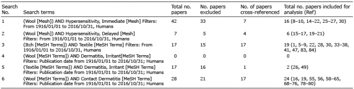
A search of Pubmed/MEDLINE was performed, and concluded in October 2016, using the following search terms in 6 separate searches (Table I); wool, textile, immediate hypersensitivity, delayed hypersensitivity, contact dermatitis, textile, irritant dermatitis. A Google Scholar search using the same key search terms was also conducted to capture relevant textile chemistry literature not present on medical databases. Articles were deemed relevant if they addressed the primary (1a and 1b) and secondary aims of the paper. Articles from the last 100 years were analyzed. Twelve papers met the primary aims and 8 met the secondary aims of the study. An additional 29 and 17 papers meeting the primary and secondary aims of the paper, respectively, were found on cross-referencing and included for analysis. To capture community perceptions of wool and allergy, a Google search was conducted using the terms wool, allergy and itch.
Table I. Search strategies and papers included for analysis
Multiple factors govern the interaction between fabric and skin that influence the wearer’s perception of garment tolerance. The primary factor is the incidence of fibre ends coarser than approximately 30–32 µm making contact with the skin (1, 2, 5–8, 22). Fibre length also influences the force exerted on the skin by the garment. Short, coarse fibres are less likely to buckle on contact with the skin and, therefore, are capable of generating sufficient force to activate afferent itch neurons (c-fibres) (1, 7). The degree of discomfort is also influenced by yarn and fabric construction, techniques to finish the fabric (2, 23), environmental factors such as the thermal environment, the integrity of the skin barrier (impaired in AD), and degree of trans-epidermal water loss (TEWL) (24). Irritation and allergy may also contribute to garment intolerance (8, 25). However, when describing cutaneous reactions to skin contact with fabrics, the term allergy is often misused in the community and not distinguished from intolerance or irritancy (9).
Irritancy describes the inflammatory cutaneous reaction following skin contact with an irritant, an agent that causes direct insult to keratinocytes (26). The chemical and physical properties of a substance determine its capacity to act as a cutaneous irritant (26). Irritant contact dermatitis is an inflammatory response of the skin following direct chemical or physical injury by an irritant (27). It results from a complex and incompletely understood interplay of both endogenous and exogenous factors activating innate immunity, without prior sensitisation (26). Since the resulting inflammation is not induced by an antigen-specific immune reaction, but through release of pro-inflammatory mediators from direct cytotoxic effects on keratinocytes (26), cutaneous irritant reactions do not represent allergy. They are, however, often associated with itch, an unpleasant sensation of the skin, provoking the desire to scratch (28). The cytotoxic release of pro-inflammatory mediators from keratinocytes, or direct physical injury caused by an irritant, can activate a distinct subpopulation of nociceptive neurons, polymodal C-fibres, that have their free nerve endings in the epidermis and dermis and are responsible for the transmission of itch (pruritoception) (24) (Fig. 1A).
Fig. 1. (A) Immune and irritant pathways contributing to the pathophysiology of atopic dermatitis. High numbers of Th2 cells (and to a lesser extent Th12 and Th22 cells) are present in AD non-lesional skin at baseline. Existing epidermal barrier protein dysfunction (i.e. filaggrin amongst others) results in increased epicutaneous penetration by allergens and irritants. Progressive activation, chemotaxis and recruitment of Th cells occurs throughout AD, but shifts in the predominant Th subsets and their associated cytokines mark the various stages. Antigen presentation by Langerhans cells (LCs) and dendritic cells (DCs) and the influence of human thymic stromal lymphoprotein (TSLP), produce a Th2 (and Th22) shift and a predominance of Th2 cytokines (interleukin (IL)-4, IL-13 and IL-31), characteristic of acute AD. Th1 and Th17 are increased to smaller extents. IL-31 activates sensory epi/dermal c-fibres leading to itch and further epidermal injury from scratching. Chronic AD is marked by significant Th1 activity (incomplete switch) as well as Th2 and Th22 intensification, producing an amplification of their associated cytokine effects. Direct keratinocyte injury from irritants (physical, chemical, thermal), leads to release of (local) pro-inflammatory mediators including the alarmin IL-33 that activates mast-cells to induce the secretion of IL-31. Adapted from: Leung & Guttman-Yassky (31) and Cevikbas et al. (38). (B) Mechanical and T-cell (T) mediated activation of cutaneous itch afferents. Mechanical activation: Fibre ends from textiles make contact with the skin. A force >75 milligraves must be exerted on the skin to activate a specific population of afferent c-fibres which transmit the itch signal to the CNS. These c-fibres express transient receptor potential (TRP) cation channels: vanilloid subtype 1 (TRPV1) and ankyrin subtype 1 (TRPA1). Fibre diameters of ≥30–32 µm are capable of generating sufficient force to activate these itch afferents. Activation is postulated to occur through mechanical triggering of TRPV1, TRPA1 and possibly also of vanilloid subtype 3 (TRPV3) and 4 (TRPV4) channels, also expressed on c-fibres and keratinocytes. T-cell mediated activation: IL-31, produced by Th2 cells predominant in AD skin, has been shown to be a central mediator of itch in AD through interaction with its receptor IL-31 receptor subtype A (IL-31RA1) that is present on a distinct population of IL-31RA1/TRPV1/TRPA1 cutaneous c-fibre and dorsal root ganglia (DRG) neurons which transmit the itch signal to the CNS. This provides a link between the T-cells predominant in AD, sensory nerves and itch. It has, therefore, been considered the so-called neuroimmune link Adapted from: Garnsworthy (5) and Cevikbas et al. (38).
On the other-hand, allergy denotes a hypersensitivity reaction mediated by antigen-specific antibodies or T-lymphocytes directed at an agent (allergen) in a previously sensitized individual (29). Cutaneous allergic reactions may be IgE-mediated (immediate or type 1 hypersensitivity, IH) or T-lymphocyte-mediated (known as delayed, or type IV hypersensitivity, DH). Exacerbations of AD can occur through both mechanisms. By contrast, contact urticaria may be mediated by IH and allergic contact dermatitis by DH.
Itch is often perceived as one of the most debilitating symptoms of allergic and atopic conditions (24), and many patients indiscriminately equate itch with allergy (8, 9, 30).
In AD, where itch is the diagnostic hallmark, the epicutaneous penetration of allergens is increased, as is the skin susceptibility to irritants (31) (Fig. 1A). Up to two-thirds of patients with AD are not atopic (29), as defined by an absence of IH following exposure to environmental allergens. The majority also do not experience DH after exposure to contact allergens (32). Therefore, their itch must be driven by other mechanisms (8, 30).
Local inflammatory mediators released from keratinocytes in response to injury/inflammation (i.e. following contact with cutaneous irritants) (33) or from leukocytic inflammation (i.e. as occurs in AD) (34), bind to specific receptors on sensory nerves to reduce pruritic activation thresholds. Direct keratinocyte injury from irritants (physical, chemical, thermal), leads to release of pro-inflammatory mediators including the alarmin IL-33 (33) and thymic stromal lymphopoietin (TSLP) (35). These activate mast cells (IL-33 and TSLP) in addition to T cells and dendritic cells (TSLP) to stimulate the secretion of interleukin (IL)-31(34). Non-neuronal acetylcholine from inflamed keratinocytes also lowers sensory nerve activation thresholds (and firing) in addition to lowering mast cell activation thresholds (36, 37).
IL-31, a known pruritus-inducing cytokine, binds to specific receptors on sensory nerves to reduce c-fibre activation thresholds via transient activation of transient receptor potential cation channels vanilloid and ankyrin subtype 1, TRPV1 and TRPA1 (34), (and possibly TRPV3 and TRPV4) (38). IL-31 produced by type 2 helper T cells (Th2) which predominate in acute AD, has been proposed as the neuro-immune link between Th2 and cutaneous itch afferents (38) (Fig. 1B). The receptor for IL-31 (IL-31RA1), exists on a distinct sub-population of cutaneous c-fibre afferents and dorsal root ganglia neurons that co-express transient receptor potential cation channel vanilloid and ankyrin subtype 1 (TRPV1 and TRPA1), capable of transmitting the itch signal to the central nervous system (24, 38) (Fig. 1B). Keratinocyte-derived TSLP (35) directly activates a subset of TRPA1-positive cutaneous sensory neurones in addition to stimulating IL-31 release by Th2 cells (35). Wilson et al. (35). demonstrated that the release of histamine or other pruritogens by mast cells was not required for TSLP-evoked itch behaviours in mice.
TRPV1 (and possibly TRPV3 and TRPV4) channels may also be responsible for transducing the force exerted on the skin by mechanical stimuli, through c-fibre activation for itch transmission (24, 38). Fibre diameters greater than 30–32 µm are capable of activating c-fibres (1, 6, 7).
Wool and other mammalian fibres, including human hair, belong to a group of fibrous proteins called alpha-keratins (39). The polypeptide chains of the wool protein consist of 18–20 amino acids (40). Hydrogen bonds between the carbonyl and amine groups of the polypeptide side chains result in protein folding into an alpha-helical arrangement (40). A characteristic feature of hard keratins such as wool, hair and nails, compared with soft keratins like those in skin, is their high proportion of disulphide bonds, formed during fibre growth by the process of keratinisation (41). These bonds stabilise the wool proteins’ tertiary structure and are responsible for the insolubility of the fibre (41).
The irritancy potential of an agent is determined by its chemical and physical properties (10, 26). For fabrics, these include mean fibre diameter, fibre length, the density of coarse protruding fibres, crimp, properties of thermal insulation and moisture transfer (6, 42). Skin penetrability contributes to an agent’s capacity to elicit irritation and is determined by molecular size, ionization state and fat solubility (26). Penetration of the stratum corneum is only achieved by low molecular weight chemicals (< 500 Daltons) (43). By contrast, the proteins comprising the wool fibre range from molecular weights of 10,000 to over 50,000 Daltons (41). Wool is also fat insoluble (41). These factors combined severely limit wool’s capacity for epidermal penetration.
The physical structural properties of fibres can cause prickle and itch, through stimulation of c-fibres (nociceptors) (1, 2). Garnsworthy et al. (1, 5) demonstrated that prickle sensation, when fabrics are worn close to the skin, is evoked by mechanical triggering of c-fibres and subsequent release of vasoactive substances. A force greater than 75 mG (milligraves) is needed to be exerted by protruding wool fibre ends to trigger C-fibres (5). The buckling load of protruding fibres is equal to the fibre diameter cubed divided by the protruding fibre length squared, so short, high diameter fibres are more likely to meet the 75 mG threshold (5). Naylor & Phillips (2), Naylor et al. (7) and Garnsworthy et al. (1, 5) showed that wool garments containing fibres with mean fibre diameters under 19 µm (7) and under 21 µm (5), were not perceived as prickly, while those with a higher incidence of fibres coarser than 32 µm caused the prickle sensation (2).
Itch triggers an efferent scratch response which attenuates itch transmission (24). It is debatable whether scratching precedes the inflammation characteristic of AD or occurs in response to inflammation (24, 44–47). However, scratching can certainly perpetuate AD. Therefore, understanding the fibre specifications responsible for c-fibre activation has important clinical relevance, particularly for patients with AD.
Correct diagnosis of allergy is dependant on a suggestive history and appropriate allergy testing, which meets certain criteria to maximise the sensitivity and specificity of the test and its interpretability (Table II). There are only 5 publications investigating whether wool is the cause of cutaneous reactions suggestive of type 1 hypersensitivity. Lord (17) reported a case of a 16-year-old girl with AD which cleared upon wool avoidance. He described dose dependant reactions to percutaneous testing (scratch or skin prick) with 1:1000 and 1:100 sheep’s wool extract and successful desensitisation. Lord also described 2 cases of acute urticaria on contact with wool in non-atopic adults. Although intradermal tests to “wool proteins” were negative, Lord (17) reproduced the urticaria on wool contact (wrapping in a woollen blanket and application of a woollen wrist cuff). In one patient, desensitization to sheep’s wool extract alleviated the symptoms. In contrast, Hill (16) conducted intradermal tests with sheep’s wool extract on a cohort of 40 children with AD and observed only borderline positive responses in 25% of patients. He interpreted these reactions to be of doubtful clinical significance (16), and hence, his results do not support immediate hypersensitivity to wool in children with AD. In 1944, Davies & Barker (15) reported 3 cases of urticaria on contact with wool in 110 admissions to the skin wards of a large military hospital with “various dermatoses”. They described patch-testing in these patients and demonstrated “skin intolerance” caused by skin contact with woollen textiles. However, it is uncertain whether these reactions represent immediate (type I) hypersensitivity to wool since patch testing is generally applied for detection of delayed (type IV) hypersensitivity and skin prick tests for measurement of specific IgE were not performed (48). These 3 studies fail to satisfy the requirements necessary for robust diagnosis of type I hypersensitivity on a number of levels, (Table III) and therefore do not provide high quality evidence in support of type 1 hypersensitivity to wool (49). None of the authors state whether negative (diluent) and positive (histamine) skin prick controls were utilised. Negative controls are necessary to exclude dermographism (50). Furthermore, the allergen extracts were not standardised or validated, and simultaneous testing of non-allergic controls was not performed raising the possibility that skin test reactions described in these studies were due to other factors, rather than to the wool fibre itself (50).
Table II. Allergy testing criteria for the accurate diagnosis of allergic disease
Table III. Summary of studies investigating cutaneous allergy to wool and their limitations according to the number of allergy testing criteria satisfied
A more recent prospective study by Wortmann et al. (52) has challenged whether wool induces cutaneous IH. Skin-prick tests (SPTs) were conducted with extracts of various types of wool buffered with solutions mimicking human sweat, (to dissolve fibre protein fractions that could potentially act as haptens for immune recognition). They demonstrated that proteins were released from both the wool fibre itself, as well as from materials used in fabric finishing; 80–97% of these proteins were smaller than 6kD (51). Three hundred and forty-eight patients presenting to the Dermatology wards of the University Hospitals in Erlangen and Bayreuth completed a questionnaire about wool (in)tolerance (51). A sub-set of 91 patients underwent SPTs by an immunologist/dermatologist using the artificial sweat buffered wool extracts (51). 70% of this group, who were predominantly atopic patients, self-identified as having wool intolerance (51). SPTs with sweat buffered blank dyed merino wool extracts in the 64 patients with self-identified wool intolerance showed no significant positive reactions relative to the negative control. Further SPT testing of other sweat buffered wool extracts (reactive dyes, afterchrome dyes and superwash – a commercially available shrink resistant treatment) generated no positive reactions (52).
Results from a study by Rasool et al. (53) also challenged the validity of IH to wool. They obtained no positive reactions in 248 patients with CU on SPT’s to a standard panel, which included sheep’s wool extract (53). The authors did not describe whether any patients had a suggestive history of CU specifically to wool. However, the negative results confirmed that wool was not a sensitiser in any of the patients tested.
No published reports of allergen-specific IgE immunoassays or SPTs to wool extracts to date have met current standards for testing; the negative study by Wortmann et al. (52) is the only study that satisfied most current testing standards, including descriptions of the major and minor constituents of the allergen extracts used, introduction of the allergen extract to the correct depth (epidermis and non-vascular superficial dermis), using appropriate SPT devices, interpretation of the reaction in the context of positive and negative controls, clear and acceptable definitions of test positivity and reporting of sufficient methodology for reproducibility. Furthermore, no specific allergenic epitopes of wool or specific IgE reacting to any such epitope have ever been identified. Wool extracts are not generally available in commercial SPT panels and data on their utility and validity is lacking.
Six studies have implicated wool as the cause of allergic contact dermatitis (14–17, 19–21). Hill (16) taped a swatch of all-white wet woollen blanket to the arm for one week to test 40 children with winter-exacerbated AD, reporting that 35% showed positive responses. Lord (17) described one case of alleged contact dermatitis occurring on sites exposed to wool. Patch testing was apparently positive to wool in this patient, however, test methodology was not described. Ramirez & Eller (20) published 4 cases of dermatitis with positive wool patch test results. They applied a swatch of wet wool to the skin that was covered by linen, rubber tissue and adhesive plaster for 24–72 h (21). The source of the swatch and its possible pre-treatment with dyes or finishes were not described. The test site was reported as clear of dermatitis and apparently healthy, although the site location was not specified (21). Davies & Barker (15) reported 10 cases with facial dermatitis, which they attributed to wool contact allergy because the dermatitis occurred after sleeping on woollen blankets. Patch tests were carried out using swatches of grey, brown and white woollen army blankets secured onto the forearm by cellophane and adhesive plaster for 48 h before reading of the results (15). Patch tests conducted with grey woollen blankets produced a severe bullous reaction, whilst those performed with brown blankets elicited only stippled erythema (15), suggesting that the reaction was secondary to the dye in the blankets rather than the wool fibre itself. Osborne & Murray (19) described 3 cases which they attributed to wool ACD, however, the history was of generalised dermatitis not confined to sites of contact with wool and patch tests were not conducted. Ballestro & Mom (14) apparently reported a case of allergic contact dermatitis to wool. However, the paper was not accessible for analysis.
Several limitations, summarised in Table III, restrict extrapolation of these study results. The study by Osborne & Murray (19) was excluded because no patch tests were undertaken. None of the studies alleging type IV hypersensitivity secondary to wool sensitisation reported the strength of the response or the criteria used to determine a positive reaction. Therefore, not only is result interpretation not standardised, but delineation of type IV hypersensitivity responses from irritant reactions is not possible. Furthermore, Hill’s (16) scraping of the arm with a tongue depressor prior to test application and the extended duration that test patches were left on the skin, in combination with the use of occlusive tapes (16, 20, 21), increased the likelihood of irritant reactions. The extent to which the obtained responses were a consequence of delayed hypersensitivity, or irritant reactions to the dyes, or finishing processes used to treat wool (55), rather than of the fibre itself, cannot be determined in these studies because patch tests were conducted with dyed or potentially treated wool/woollen blankets. Therefore, cross-contamination of allergens may have confounded the results obtained (54).
Textile allergy (ACD, CU) and irritant contact dermatitis (27, 55) to various dyes and finishes used in garment manufacturing (rather than to wool fibre itself) are well described (56). Formaldehyde is used widely in many consumer products including resins for pressed wood products, housing insulation, and adhesives, in addition to use in low concentrations in cosmetics, cleaning products and textiles (57). ACD from formaldehyde, used in the textile industry since the 1920s to prevent wrinkling, has been documented for many fibre types (58, 59). Hellier (60) and Romaguera et al. (61) suggested that purpuric eruptions associated with wool could also be secondary to allergy from a urea or melamine-formaldehyde resin finish. The degree of reported formaldehyde sensitisation from clothing has varied from 11% to 36% in published studies (62–64). Up to 83% sensitisation was reported by Burry et al. (65). Although formaldehyde has historically been used widely in clothing (particularly cotton) as well as in cosmetic products, a 2007 Australian Competition and Consumer Commission (ACCC) study of a broad range of clothing purchased in the Australian market detected no residual formaldehyde in woollen or cotton garments (57). The ACCC have adopted interim non-regulatory maximum residue benchmarks for formaldehyde in textiles (66). By comparison, China and Japan have legally limited clothing formaldehyde levels. The United States of America (USA) have no legal limit and have recently found formaldehyde in various cotton and cotton/synthetic mixes, at levels (70–206 ppm) well exceeding the 30ppm thought capable of sensitization, but not in wool (67). People sensitised to formaldehyde are recommended fabrics such as 100% wool by the ACCC, as these are unlikely to have been treated with formaldehyde resins (57). Formaldehyde contact allergy is therefore no longer a concern for modern woollen garments (68).
Chrome dyes, used mainly in the dyeing of wool and silk to darker colours, require after treatment with a mordant (potassium dichromate). Five cases of ACD were described in serviceman from chromium present in green wool uniforms (69, 70). However, chrome dyes are decreasingly used, mainly for ecological reasons. New processes with iron-complexed after-chrome dyes have reduced chromium in dye effluents whilst maintaining colour fastness (71, 72). There have been no reports of IH to chrome dyes in textiles. Although chromium is a well known contact allergen, fabrics have not been a source of sensitisation (73). A literature review of textile dye dermatitis by Hatch & Maibach (74) similarly reported no cases of ACD owing to after-chrome dyes. The absence of reports of allergic cutaneous reactions from after-chrome dyed textiles since 1978 may suggest that modern after-chrome dyes are poor sensitisers. Conversely, disperse dyes (particularly blue 124 and orange 3), used to colour polyester, acetate and nylon fibres, are now the most common textile dye sensitizers (75). The overall rate of sensitisation to reactive dyes used to colour natural fibres including cotton silk and wool is very low: 18 of 1,813 patients (0.99%) tested with the additional textile series had positive reactions to reactive dyes over 1 year (76). Of the reactive dyes used to colour natural fibres, Cibracron Cr and Violet Remazol 5R were found to be the commonest sensitisers (8/18 and 5/18 positive PT’s respectively) (76).
Lanolin originates from wool wax (sheep sebum) and is composed of free fatty alcohols, esters and hydroxyl esters of long-chained alcohols (aliphatic alcohols, sterols), and fatty acids. It is added to many consumer products, particularly cosmetics (68). Ramirez & Eller (21) documented the first lanolin patch-test-positive case of ACD. However, today’s wool scouring systems remove most of the lanolin from wool to levels less than 0.5%, since higher levels can reduce fabric quality (77). Subsequent dyeing and finishing operations reduce residual lanolin levels even further. Although previously considered an important sensitiser (78, 79), recent data does not support this. The mean annual rate of lanolin sensitivity was found to be 1.7% on retrospective database review of 24,449 patients patch tested between 1982–1996 with a standard series containing 30% wool alcohols in a central London teaching hospital (80). Therefore, the likelihood of lanolin contact dermatitis occurring as a result of wearing wool is very low for modern garments.
Given wool and human hair belong to the same group of alpha-keratin proteins and therefore share may properties, evidence for type I or IV hypersensitivity to human hair was examined. A study by Uehara & Ofuji (81) reported that almost two thirds of patients with AD exhibited contact hypersensitivity to human dander, with positive patch tests obtained in 66% of patients with AD compared with 3% of normal controls. However, the patch test methods employed by Uehara & Ofuji (81) fail to meet today’s standards, and therefore, make delineation of type IV hypersensitivity from irritant reactions difficult in this study. More robust patch testing methodology and interpretation is described by Sasaki et al. (82), who reported positive patch tests for the acetone soluble fraction of human dander in 38% of patients with AD, whereas reactions were rare in normal and clinical controls, suggesting an increased incidence of type IV contact hypersensitivity to human dander in AD. However, the scarcity of cases published on contact dermatitis to human hair, in addition to the lack of methodological detail reported, limit the interpretability of the results and their extrapolation to wool (20, 21).
To date there is an absence of evidence to substantiate allergy (type I and IV hypersensitivity) to wool fibres. Furthermore, allergens associated with wool processing (eg chemical dyes) are present at negligible levels within modern wool garments. Mean fibre-diameter in particular is closely linked to perceptions of garment irritancy. Coarse fibres – whether from wool or other fabrics – are able to cause cutaneous irritancy, whereas fabrics with finer fibre diameters, such as superfine and ultrafine Merino wool, are not perceived as irritating.
Research over the last decade has uncovered a number of new pruritogenic mediators beyond histamine (24). Detailed review of their role in pruritus is beyond the scope of this paper. However, it is important to recognise that although histamine may be released by allergen-specific IgE degranulation of mast cells, mast cell activation and keratinocyte histamine release also occur through a number of non-allergic mechanisms (83). Histamine activates mechanically-insensitive afferent (MIAs) c-fibres involved in itch transmission (84). Some of these newly recognized mediators are thought to utilize this pathway for the transmission of itch. However, human electrophysiological studies have demonstrated the existence of a second peripheral pathway for itch that can be activated by non-histaminergic pruritogens (84). Moreover, thermal, chemical and mechanical cutaneous stimuli can directly activate sensory nerve endings and/or keratinocytes via a family of transient receptor potential (TRP) channels (24). Activation of TRPV1, TRPV3, TRPV4 and TRPA1 channels, expressed in human keratinocytes and free cutaneous sensory nerve endings, transduce signals to the dorsal root-ganglion (DRG) (24). These mechanisms may represent a plausible pathway for wool fibres to induce itch through exertion of a mechanical cutaneous stimulus, which is independent of allergy. Based on results from Garnsworthy et al. (1) and Naylor et al. (7), the fibres of superfine and ultrafine Merino wool garments, with mean fibre diameters of 15–18.5 µm and 11.5–15 µm respectively, would not generate the mechanical force required to induce mechanical activation of nociceptors, Hence, superfine and ultrafine Merino wool garments may not be capable of generating sufficient incidence of TRP channel activation, therefore allaying the stimulus for itch, intolerance and irritancy.
Recent Australian studies have demonstrated that the wearing of superfine Merino wool garments next to the skin may significantly reduce AD severity in children under 3 years with mild AD (85) and in persons aged between 6–81 years of age with mild-severe AD (86). In these studies, the superfine Merino wool fibre did not produce cutaneous irritation in patients with AD. The mechanism responsible for the potentially therapeutic benefit of superfine Merino wool in these patients is unclear, but may relate to micro-climate optimization when worn next to the skin.
The evidence to date fails to support the notion that wool is an allergen, or that the wool fibre causes cutaneous allergic reactions (mediated by either type I or IV hypersensitivity). The papers implicating wool as the cause of cutaneous allergic reactions have important limitations that counter their findings. In fact, studies with fewer limitations and stronger SPT methodology demonstrate no evidence of type I HS to wool. More detailed characterisation of wool components with potential to act as allergenic epitopes (not identified to date) and investigation of allergenic specificities of IgE directed against wool components would be necessary to completely exclude the possibility of wool allergy. However, findings to date highly dispute the plausibility of wool allergy and therefore, limit incentives for research into allergenic epitopes or IgE specificities against wool.
Cutaneous irritation caused by wool garments in previous years was due to high fibre diameters. Increasingly available superfine and ultrafine Merino wool garments with finer fibre diameters do not activate sufficient c-fibres responsible for itch transmission and are well tolerated (87). Coarse diameter fibres of any fabric may irritate, but irritation is neither a general nor a specific property of wool. In fact, recent studies suggest that superfine or ultrafine Merino wool with finer fibre diameters may benefit AD management (85). Known allergens associated with textile processing are minimally present in wool garments today given current industry practices and are unlikely to lead to allergic reactions. Further studies examining the potential benefit of superfine wool in AD management, the therapeutic mechanism for such a benefit and formal patch testing studies with superfine wool would be beneficial.
The authors declare no conflicts of interest.


 Click to show fullsize
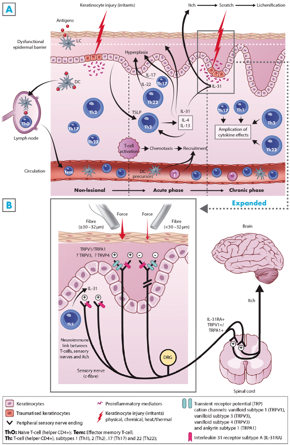 Click to show fullsize
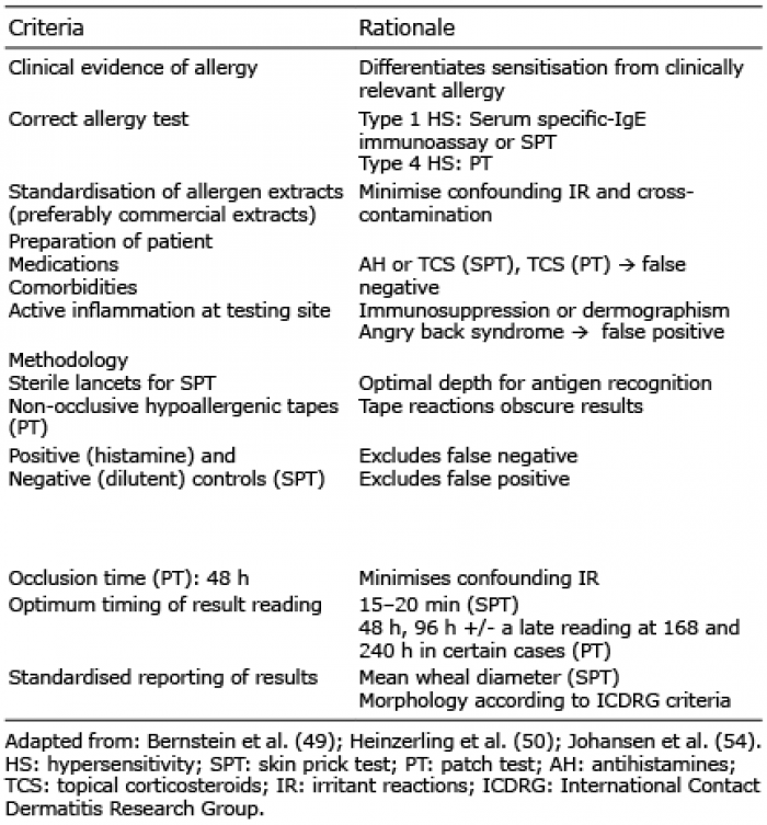 Click to show fullsize
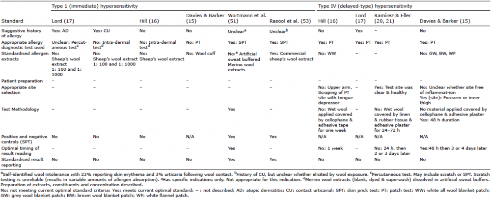 Click to show fullsize