


Noah Scheinfeld
Department of Dermatology, St Luke’s Roosevelt Hospital Center, New York, USA
Neoplastic cells, both malignant and benign, local occurring and metastatic, can cause alopecia of the scalp. However, the infiltration of neoplastic cells is sometimes not florid; a condition known as “scalp alopecia due to a clinically unapparent or minimally apparent neoplasm” (SACUMAN). Neoplastic cells can nevertheless destroy hair follicles by inducing fibroplasias via inflammatory mediators, attracting inflammatory cells and/or replacing normal cellular populations. The infiltrative nature of such an alopecia can be unapparent or only minimally apparent. The most common neoplasm in which an uncomplicated, minimally or unapparent scalp alopecia occurs and no infiltrate of cancer is suspected is metastatic breast carcinoma. Other causes include squamous and basal cell carcinomas, angiosarcoma, gastric carcinoma, placental site tromphoblastic tumor, and mycosis fungoides. Syringoma-like proliferations can underlie alopecia. It is unclear whether these proliferations are true syringomas or normal findings. In conclusion, neoplasms causing cicatricial alopecia of the scalp are very rare, so generalizations from the limited number of case reports are of uncertain importance. Moreover, it is likely that many cases of neoplasms causing cicatricial alopecia of the scalp are diagnosed as inflammatory alopecia and not neoplasms, thus depriving us of a full accounting and understanding of this entity. Dermatologists must be aware that in rare cases a bland scalp alopecia can represent a new or recurring, local or metastatic neoplasm. Key words: alopecia; alopecia neoplastica; breast cancer; neoplasm; cicatricial alopecia.
(Accepted July 25, 2006.)
Acta Derm Venereol 2006; 86: 387–392.
Noah Scheinfeld MD, Department of Dermatology, St Luke’s Roosevelt Hospital Center, 1090 Amsterdam Ave, New York NY 10025, USA. E-mail: Scheinfeld@earthlink.net
Scalp alopecia has a variety of causes. Alopecia can be due to factors that primarily and intrinsically affect the follicles, as in androgenic alopecia. Alopecia can also be due to conditions and factors that extrinsically and secondarily affect the follicles. Secondary types of alopecia are caused by inflammatory diseases (e.g. discoid lupus), trauma, infections, and infiltration of neoplastic cells (1–9). Sometimes keloids can cause alopecia in striking fashion and be mistaken for tumors (Fig. 1). Sometimes the infiltration of neoplastic cells manifests as papules and nodules and a diagnosis of cutaneous metastasis can be made clinically. On other occasions the infiltration of neoplastic cells is unapparent or minimally apparent on physical examination. In the latter, clinically an inflammatory or hormonal alopecia is suspected, a suspicion that is overturned as the cancer expands it physical manifestations. This paper will review such scalp alopecia (due to a) clinically unapparent or minimally apparent neoplasm (SACUMAN) (Table I).
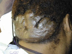
Fig. 1. Keloid mimicking tumor on the back of the neck, with total alopecia in the keloid area.
Table I. Various causes of alopecia neoplastica of the scalp without clinically apparent neoplasm at the site of alopecia (SACUMAN)
| Metastatic malignant neoplasms | Locally occurring malignant neoplasms | Benign neoplasms |
| Breast cancer Gastric cancer Placental site trophoblastic tumor | Basal cell carcinoma Melanoma Angiosarcoma Mycosis fungoides Syringomatous carcinoma Proliferating trichilemmal tumors Squamous cell carcinoma Plasmacytoma Mastocytosis | Syringomas Nerve sheath myxoma Nevocellular nevus Neuroma Trichoepithelioma Osteomyoma-like tumor |
Mechanism of action
The precise mechanisms causing SACUMAN are not fully defined (5). Neoplastic cellular infiltration, by definition, has not fully overrun the normal cellular population (otherwise it would be visible to the naked eye) and thus does not fully explain the destruction of the pilosebaceous unit. Histopathologically, there are usually neoplastic cells in a dense dermal and subcutaneous collagenous stroma without pilosebaceous units. There is often dermal fibroplasia. The scarring that breast cancer induces is not irreversible after it manifests, suggesting some alopecia-inducing mechanism independent of mere fibroplasias, as sometimes alopecia induced by metastatic breast cancer can be accompanied by scalp hair regrowth after effective cancer treatment (9).
Cytokine and interleukin production by neoplastic cells may explain SACUMAN. In inflammatory types of alopecia, the pattern of cytokine expression shows the presence of fibrogenic molecules (interleukins 4 (IL-4) and 6 (IL-6), basic fibroblast growth factor (bFGF) and transforming growth factor-beta (TGF-β)) (2). TGF-β1 promotes breast cancer progression and measurement of tumoral TGF-β1 levels, especially for node-negative patients, identifies a high-risk population early in breast cancer progression (3). Thus, the TGF-β1 secreted by the breast cancer may cause the scalp alopecia that sometimes accompanies breast cancer. Patients with head and neck squamous cell carcinoma, which can be associated with alopecia of the head and neck, have increased levels of IL-4 (4), an interleukin associated with alopecia.
Crotty et al. (5) postulated that the release of cytokines from tumor cells caused the disappearance of the hair follicle. They noted a case in which infiltration of the hair follicles by melanoma cells was clearly present and only a mild inflammatory component was seen. Rather than inflammatory cells being the only motive factor behind neoplastic alopecia, Crotty et al. (5) suggested that cytokines released from neoplastic and fibrous tissue, in particular bFGF, caused alopecia. That is, malignant tumors evoked a stromal response manifesting as fibroplasia lacking follicular structures. Others too noted fibrosis associated with alopecia due to neoplastic cells (6, 7). Murray et al. (8) speculated that direct effects of androgenic or other hormonal stimuli caused alopecia, although little histological evidence supports this suggestion. While much work needs to be done to define the interleukin and cytokine profiles of neoplastic and inflammatory alopecia, it is possible that they share common cytokine and interleukin profiles.
THE ETIOLOGIC SPECTRUM
The underlying causes of SACUMAN are summarized in Table I. They can be classified as metastatic or locally occurring (malignant or benign) neoplastics.
Breast cancer
The most common cause of SACUMAN is breast cancer (9–13). This alopecia has a variety of degrees of severity. Mallon & Dawber (14) reported a patient with breast carcinoma in whom hair loss was clinically inconspicuous. Breast cancer causing SACUMAN is often described as alopecia areata-like. This is a misnomer because alopecia neoplastica is a cicatricial process (13–19). The severity of the alopecia induced by breast cancer can be severe and has been reported to manifest as total scalp alopecia accompanied by alopecia totalis (10). As stated, sometimes alopecia induced by metastatic breast cancer will remit with effective cancer treatment showing that the scarring process, once under way, is reversible if not complete (9).
Non-breast carcinomas
SACUMAN can be due to internal cancers other than breast carcinoma. Kim et al. (7) reported a 36-year-old woman with an asymptomatic erythematous alopecic plaque that had developed 10 months before presentation of a gastric carcinoma with pelvic metastases. Yuen et al. (18) described a 31-year-old woman who had been pregnant and given birth three times who presented with several alopecic patches resembling alopecia areata, which on biopsy proved to be metastatic, previously unsuspected, placental site trophoblastic tumor. Again while these processes are described as alopecia areata like this is a misnomer because biopsies reveal a scarring process.
Lymphomas
While B-cell lymphoma can cause cutaneous tumors in the scalp, these tumors do not cause SACUMAN (20). Mycosis fungoides (MF) (also known as cutaneous T-cell lymphoma (CTCL)), however, can be associated with SACUMAN due to both infiltration of neoplastic T cells and its common clinical manifestation as follicular mucinosis (21–25). MF most commonly causes alopecia due to follicular mucinosis (5–7). In this condition, intra-follicular mucin deposition results in follicle destruction and hair loss. A discussion of follicular mucinosis is beyond the scope of this paper, however, it is important to note here that sometimes this condition is not clinically apparent.
Follicular mucinosis is not the only mechanism by which MF causes alopecia. Infiltration of the neoplastic T cells into the skin of the scalp can result in alopecia. Kossard et al. (22) reported a patient with MF who developed extensive alopecia. Multiple scalp biopsies showed perifollicular and intrafollicular infiltrate of lymphocytes, but no follicular mucinosis. On transverse sections many of the follicles lacked hair sheath, canal, sebaceous gland or hair differentiation and instead formed undifferentiated basaloid structures, resembling cutaneous lymphadenoma. The alopecia of CTCL can overlie areas of extensive cerebral bone destruction (23). Syringolymphoid hyperplasia of the scalp can be associated with alopecia and some believe that syringolymphoid hyperplasia with alopecia is in fact CTCL (24).
Panniculitis-like T-cell lymphoma is a condition distinct from MF, which can clinically manifest as alopecia. Torok et al. (25) reported a 45-year-old woman who presented with multifocal scalp lesions with the clinical impression of alopecia areata whose histological findings first suggested cytophagic histiocytic panniculitis, although a “burned-out” panniculitis-like T-cell lymphoma could not be excluded; after a 20-month follow-up period, assessment of the T-cell receptor gamma-chain gene rearrangement supported the diagnosis of subcutaneous panniculitis-like T-cell lymphoma. It should, of course, be noted that the presence of clonal rearrangement does not necessarily equate with lymphoma.
Basal cell carcinoma
Basal cell carcinoma (BCC) can cause SACUMAN (26), unsurprisingly because BCC has protean clinical presentations (27). Cicatricial alopecia of the scalp caused by BCC can mimic alopecia areata and manifest without the crusted plaques or papules or nodules with pearly borders (28). Alopecia of any sort associated with BCC, however, is a rare entity. In a series compiled by Labareda & Garcia e Silva (29) of 3000 BCCs, 77 were on the scalp with the following significant associated factors noted in 19 cases: organoid nevi (11), syringocystadenoma papilliferum (1), androgenic alopecia (5), wound scar (1) and radiodermatitis (1). It is not clear from this article whether these latter 3 categories were SACUMAN-related to BCC or other forms of alopecia co-incident to a BCC.
Melanocytic neoplasms
Malignant and benign melanocytic processes can mimic, be hidden by or induce alopecia in the scalp (30). Melanoma can be related to alopecia. Crotty et al. (5) reported a 50-year-old man presenting with a 6-month history of localized alopecia on the left frontal area of the scalp, whose biopsy revealed desmoplastic melanoma with associated neurotropism in which hair follicles were focally reduced in number and were infiltrated by melanoma cells. Melanoma can occur in areas of alopecia without mimicking alopecia or without occurring in an occult fashion.
Nevi can also be related to alopecia (31). Nevocellular nevus associated with alopecia presenting as aplasia cutis congenita has been reported (32). Alopecia that histologically resembled a halo nevi with an inflammatory infiltrate concentrated around the hair follicles rather than the nevus has been reported (33).
Angiosarcoma
Cutaneous angiosarcoma can present as SACUMAN (Fig. 2) (34). Murray et al. (7) reported an 83-year-old woman presenting with a 6-month history of hair loss and painless bruising involving her forehead and scalp whose scalp biopsy revealed angiosarcoma with a significant increase in miniaturized and telogen hair follicles and some tumor-associated scarring hair loss present only in areas of tumor involvement and not in a typical distribution pattern for androgenetic alopecia.
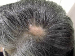
Fig. 2. Circumscribed circular area of scalp alopecia overlying angiosarcoma.
Neural proliferations
Proliferations of neural tissue can cause or underlie SACUMAN. Hernandez-Cano et al. (35) reported patchy alopecia areata-like hair loss around a solitary circumscribed neuroma. Burket (36) described a 16-year-old girl with a 2.5-cm macular area of alopecia of the posterior portion of the scalp in which a biopsy revealed a subcutaneous nerve sheath myxoma; hair growth resumed 4 weeks after its surgical removal. Neural tissue of various sorts can underlie alopecia, in particular in neonates (37).
Hematologic processes
Pathology of mast cells (38) and plasma cells (39) involving the skin has been related to SACUMAN. Weichenthal et al. (40) described a male with a 2-year history of alopecia, cervical lymphadenopathy, erythematous thickening of the skin on the neck, and progressive walking difficulties. A plasmacytoma with lambda cell restriction was found. The overlying skin showed marked fibrosis, with loss of hair follicles, and a plasma cell infiltrate of polyclonal origin. Cervical lymph nodes showed multicentric Castleman’s disease. After excision of the plasmacytoma and postoperative irradiation, the alopecia remained but did not progress.
Mischellaneous tumors of local origin
Syringomatous carcinoma, proliferating trichilemmal tumor and squamous cell carcinoma have been reported as causes of SACUMAN. Alessi & Caputo (41) reported a case of well-differentiated syringomatous carcinoma in a 50-year-old man. The syringomatous carcinoma was located on the scalp and manifested as a slowly enlarging patch of alopecia misdiagnosed as alopecia areata for years after erroneously and initially having been considered, based on a biopsy, to be a benign syringoma. This cancer was not unapparent, yet its bland and slight appearance did raise suspicion in the patient’s physicians that he had a cancer. Dekio et al. (42) reported a 43-year-old man with a proliferating trichilemmal tumor that appeared on an area of pre-existing alopecia underlined by ectopic apocrine sweat glands. Squamous (spinous) cell carcinoma has occurred in association with SACUMAN (43).
Benign neoplasms causing cicatricial alopecia of the scalp
Benign neoplastic proliferations have been linked with SACUMAN. These include: trichoepitheliomas, osteoma like proliferations and, most commonly, syringomas, and syringoma-like proliferations.
Rare reports link benign neoplasms besides syringomas with SACUMAN. A 13-year-old girl had a 6-year history of facial infiltrated erythematous plaques, alopecia of the eyebrows, diffuse alopecia of the scalp, and absence of body hair, simulating lepromatous leprosy, which, histologically, were trichoepitheliomas (44). Alopecia with fibrous dysplasia manifesting as a strange coil of fibrous tissue and osteomas, histologically analogous to that occurring within the bones in Albright disease, has been noted in patients with polyostotic fibrous dysplasia (45).
The hair is associated intimately with eccrine glands. Therefore, it is unsurprising that pathologic changes in the eccrine glands can adversely affect hair follicles. Syringomas and syringoma-like eccrine sweat duct proliferation can underlie SACUMAN (Fig. 3). Based on the author’s personal experience it seems that some of these proliferations of eccrine ducts are incidental findings that are not the primary cause of the alopecia that overlies such proliferations. Consonant with this observation, Mehregan & Mehregan (46), who reviewed a series of 585 scalp biopsies taken for histologic evaluation of hair loss and noted syringoma-like eccrine sweat duct proliferation in 6 specimens with scarring alopecia and one each with alopecia areata and female-pattern alopecia, believed that eccrine overgrowth did not have a major role in causing alopecia. A number of authorities disagree with these authors and believe that eccrine sweat duct hamartomas (47) and syringomas cause scarring alopecia (48–51), progressive hair loss (52), and alopecia with a sclerotic quality (53).

Fig. 3. Circumscribed circular area of scalp alopecia overlying syringoma-like eccrine sweat duct proliferation.
CONCLUSION
SACUMAN must be recognized as a rare and relevant cause of hair loss. It is important to recognize that SACUMAN exists because patient with hair loss and no or minimal scalp surface change, who are in certain risk groups (e.g. women with a history of cancer) might need actual scalp biopsies to document the cause of their alopecia. Failing to follow on scalp alopecia that somehow does not fit the pattern of androgenic alopecia seems a particularly unnecessary source of morbidity and mortality. SACUMAN’s most common cause is metastatic breast carcinoma (54–56). When the writers of case reports describe cicatricial alopecia of the scalp due to neoplasms as alopecia areata-like, which is the most common description of cicatricial alopecia of the scalp due to neoplasms, it is a misnomer. Isolated plaques of scalp alopecia should prompt investigation. As Crotty et al. (5) aptly state: “a scarring alopecia that is not readily explained by trauma, infection or dermatosis should lead to the consideration of an underlying neoplastic process. Biopsy is required. The presence of associated neurological signs should also suggest neural involvement by a neoplastic process.” The mechanisms behind the development of SACUMAN require further definition, but might involve TGF-β and IL-4. An awareness of SACUMAN assists the accurate diagnosis of patients with scalp hair loss and underlying neoplasms.
Acknowledgement
The author thanks Richard Vinson for providing Fig. 2.
References
1. Baum EM, Omura EF, Payne RR, Little WP. Alopecia neoplastica – a rare form of cutaneous metastasis. J Am Acad Dermatol 1981; 4: 688–694.
2. Moretti S, Amato L, Massi D, Bianchi B, Gallerani I, Fabbri P. Evaluation of inflammatory infiltrate and fibrogenic cytokines in pseudopelade of Brocq suggests the involvement of T-helper 2 and 3 cytokines. Br J Dermatol 2004; 151: 84–90.
3. Desruisseau S, Palmari J, Giusti C, Romain S, Martin PM, Berthois Y. Determination of TGFbeta1 protein level in human primary breast cancers and its relationship with survival. Br J Cancer 2006; 94: 239–246.
4. Lathers DM, Young MR. Increased aberrance of cytokine expression in plasma of patients with more advanced squamous cell carcinoma of the head and neck. Cytokine 2004; 25: 220–228.
5. Crotty K, McCarthy W, Quinn M, McCarthy S. Alopecia neoplastica caused by desmoplastic melanoma. Australas J Dermatol 2003; 44: 295–298.
6. Cohen I, Levy E, Schreiber H. Alopecia neoplastica due to breast carcinoma. Arch Dermatol 1961; 84: 490–492.
7. Kim HJ, Min HG, Lee ES. Alopecia neoplastica in a patient with gastric carcinoma. Br J Dermatol 1999; 141: 1122–1124.
8. Murray S, Simmons I, James C. Cutaneous angiosarcoma of the face and scalp presenting as alopecia. Australas J Dermatol 2003; 44: 273–276.
9. Archer CB, Smith NP. Alopecia neoplastica responsive to tamoxifen. J R Soc Med 1990; 83: 647–648.
10. Martin J, Ross JB. Alopecia totalis as a presentation of cutaneous metastasis (alopecia neoplastica). Int J Dermatol 1983; 22: 487–489.
11. Friedmann E. Metastatic epithelioma of the scalp, probably of mammary origin; primary infiltrating alopecic type. Bull Soc Fr Dermatol Syphiligr 1969; 76: 453–454.
12. Vilmer C, Trassard M. A case for diagnosis: cutaneous metastasis of breast carcinoma. Ann Dermatol Venereol 1993; 120: 561–562.
13. Canas R, Vidarte, Navarrete. Sclerothrophic alopecias by breast carcinoma metastasis Actas Dermosifiliogr 1979; 70: 243–248.
14. Mallon E, Dawber RP. Alopecia neoplastica without alopecia: a unique presentation of breast carcinoma scalp metastasis. J Am Acad Dermatol 1994; 31: 319–321.
15. Carson HJ, Pellettiere EV, Lack E. Alopecia neoplastica simulating alopecia areata and antedating the detection of primary breast carcinoma. J Cutan Pathol 1994; 21: 67–70.
16. Schorr WF, Swanson PM, Gomez F, Reyes CN. Alopecia neoplastica. Hair loss resembling alopecia areata caused by metastatic breast cancer. JAMA 1970; 213: 1335–1337.
17. Haas N, Hauptmann S. Alopecia neoplastica due to metastatic breast carcinoma vs. extramammary Paget’s disease: mimicry in epidermotropic carcinoma. J Eur Acad Dermatol Venereol 2004; 18: 708–710.
18. Yuen YF, Lewis EJ, Larson JT, Wilke MS, Rest EB, Zachary CB. Scalp metastases mimicking alopecia areata. First case report of placental site trophoblastic tumor presenting as cutaneous metastasis. Dermatol Surg 1998; 24: 587–591.
19. Schultz-Ehrenburg U, Thies W. Alopecia scleratrophicans carcinomatosa in metastasizing carcinoma of the breast. Z Hautkr 1975; 50: 141–146.
20. Magina S, Mesquita-Guimaraes J, Resende C, Bello M, Dias C. Scalp tumour as a sign of systemic B-cell lymphoma. J Eur Acad Dermatol Venereol 2004; 18: 196–198.
21. Jackow CM, Papadopoulos E, Nelson B, Tschen JA,
Heatherington G, Duvic M. Follicular mucinosis associated with scarring alopecia, oligoclonal T-cell receptor V beta expansion, and Staphylococcus aureus: when does follicular mucinosis become mycosis fungoides? J Am Acad Dermatol 1997; 37: 828–831.
22. Kossard S, White A, Killingsworth M. Basaloid folliculolymphoid hyperplasia with alopecia as an expression of mycosis fungoides (CTCL). J Cutan Pathol 1995; 22: 466–471.
23. Natsuda H, Muraki Y, Kobayashi T, Kozima H, Shibuya A, Nagasawa T, Abe T. Adult T cell lymphoma/leukemia with alopecia, huge tumors of scalp and wide destruction of cerebral bone Rinsho Ketsueki 1991; 32: 537–541.
24. Esche C, Sander CA, Zumdick M, Kutzner H, Kind P, Schulte K, et al. Further evidence that syringolymphoid hyperplasia with alopecia is a cutaneous T-cell lymphoma. Arch Dermatol 1998; 134: 753–754.
25. Torok L, Gurbity TP, Kirschner A, Krenacs L. Panniculitis-like T-cell lymphoma clinically manifested as alopecia. Br J Dermatol 2002; 147: 785–788.
26. Schmid MA. Basalioma of the scalp. Langenbecks Arch Chir 1968; 320: 155–178.
27. Fitzpatrick JE, Whalen EA. Basal cell carcinoma or not? Histological variants and mimics of the most common cutaneous malignancy. Semin Cutan Med Surg 1999; 18: 15–24.
28. Hundeiker M, Tillmann U. Basal cell carcinomas of the hairy head simulating “alopecia areata.” Hautarzt 1972; 23: 180–181.
29. Labareda JM, Garcia e Silva L. Basal cell carcinomas of the scalp. Review of 77 patients with 81 tumors. Med Cutan Ibero Lat Am 1988; 16: 367–372.
30. Bricout N, Raulo Y, Geissmann O, Wechsler J. Association of a melanoma and a spinocellular epithelioma on a burn scar of the scalp. Sem Hop 1983; 59: 2933–2935.
31. McDonagh AJ, Laing RW, Harrington CI, Griffiths RW. Giant alopecic nodule of the scalp: unusual presentation of a cellular blue naevus in an adult. Br J Dermatol 1992; 126: 375–377.
32. Mastruserio DN, Cobb MA, Ross VE. Nevocellular nevus associated with alopecia presenting as aplasia cutis congenita. Int J Dermatol 1998; 37: 37–39.
33. Yesudian P, Thambiah AS. Perinevoid alopecia. An unusual variant of alopecia areata. Arch. Dermatol 1976; 112: 1432–1434.
34. Knight TE, Robinson HM Jr, Sina B. Angiosarcoma (angioendothelioma) of the scalp. An unusual case of scarring alopecia. Arch Dermatol 1980; 116: 683–686.
35. Hernandez-Cano N, Pizarro A, Lazaro TE, Mayor M, Buron I, Contreras F, Casado M. Nonscarring alopecia associated with solitary circumscribed neuroma. Dermatology 1997; 195: 265–267.
36. Burket JM. Alopecia associated with underlying nerve
sheath myxoma. J Am Acad Dermatol 1987; 16: 209–211.
37. Rogers GF, Mulliken JB, Kozakewich HP. Heterotopic neural nodules of the scalp. Plast Reconstr Surg 2005; 115: 376–382.
38. Bureau Y, Barriere H, Litoux P, Bureau B. Mastocytosis in a brother and sister, with pseudopeladic localization in the scalp, in the sister. Bull Soc Fr Dermatol Syphiligr 1971; 78: 240–241.
39. Janner M, Lippert HD, Stolzenbach G. Follicular mucinosis and large area, partly lichenoid, partly sclerodermiform generalized paramyloidosis as a cutaneous paraneoplastic syndrome in myeloma (IgD and light chain plasmacytoma). Z Hautkr 1974; 49: 673–681.
40. Weichenthal M, Stemm AV, Ramsauer J, Mensing H,
Feller AC, Meigel W. POEMS syndrome: cicatricial alopecia as an unusual cutaneous manifestation associated with an underlying plasmacytoma. J Am Acad Dermatol 1999; 40: 808–812.
41. Alessi E, Caputo R. Syringomatous carcinoma of the scalp presenting as a slowly enlarging patch of alopecia. Am J Dermatopathol 1993; 15: 503–505.
42. Dekio S, Imaoka C, Jidoi J. Proliferating trichilemmal tumor with apocrine sweat glands. J Dermatol 1990; 17: 391–393.
43. Lliteras JV, Cabre J. Spinous cell carcinoma on erythematode cicatricial alopecia. Actas Dermosifiliogr 1971; 62: 63–68.
44. de la Luz Orozco-Covarrubias M, Uribe-Rea C, Tamayo-Sanchez L, Duran-McKinster C, Ruiz-Maldonado R. Trichoepitheliomatous infiltration of the skin simulating leprosy. Pediatr Dermatol 1993; 10: 252–255.
45. Shelley WB, Wood MG. Alopecia with fibrous dysplasia and osteomas of skin. A sign of polyostotic fibrous dysplasia. Arch Dermatol 1976; 112: 715–719.
46. Mehregan AH, Mehregan DA. Syringoma-like sweat duct proliferation in scalp alopecias. J Cutan Pathol 1990; 17: 355–357.
47. Trozak DJ, Wood C. Occult eccrine sweat duct hamartoma and cicatricial scalp alopecia. Cutis 1984; 34: 475–477.
48. Helm TN, Guitart J, Bergfeld WF, Benedetto E. Occult syringoma associated with alopecia. Int J Dermatol 1992; 31: 437–438.
49. Neuman KM, Burnett JW. Alopecia associated with
syringomas. J Am Acad Dermatol 1985; 13: 528–529.
50. Tsoitis G, Asvesti C, Mandinaos C, Lambroudi M, Hatzibougias J, Babi A. Cicatricial alopecia of the scalp with histological aspect of syringoma. Ann Dermatol Venereol 1992; 119: 919–922.
51. Dupre A, Bonafe JL, Christol B. Syringomas as a causative factor for cicatricial alopecia. Arch Dermatol 1981; 117: 315.
52. Shelley WB, Wood MG. Occult syringomas of scalp associated with progressive hair loss. Arch Dermatol 1980; 116: 843–844.
53. Noble JP, Lessana-Leibowitch M, Sedel D, Guillemette J,
Cadot M, Hewitt J. Scalp syringoma with patch of alopecia, lichen sclerosus. Ann Dermatol Venereol 1979; 106: 275–277.
54. Velez JR, Ferrando J, Palou J, Mascaro JM. Metastatic alopecia. Med Cutan Ibero Lat Am 1990; 18: 185–188.
55. Hernanz JM, Vives P, Garcia Almagro D, Jaqueti G. Neoplastic alopecia. Actas Dermosifiliogr 1979; 70: 507–514.
56. Camps Freneda A, Boleda M, Cerdan M, Mendez R, Debray N. Metastatic alopecia. Actas Dermosifiliogr 1979; 70: 313–317.

