


1Coeliac Disease Research Center, Faculty of Medicine and Life Sciences and 4Faculty of Social Sciences, University of Tampere, Departments of 2Dermatology, 3Gastroenterology and Alimentary Tract Surgery, and 5Internal Medicine, Tampere University Hospital, Tampere, Finland
Coeliac disease and dermatitis herpetiformis (DH) are characterized by autoantibodies targeting transglutaminase (TG)2 and TG3, respectively. Previous studies show that TG2 antibodies are produced in the gut and can be assessed in organ culture of small-intestinal biopsies from patients with coeliac disease. Thus far, no studies have investigated TG3 antibodies in organ culture of biopsies from patients with DH, or exploited the method in DH. The aim of this study was to investigate TG3 and TG2 antibody responses in serum and small-intestinal biopsies from patients with DH with active disease, and from those in remission. The majority of patients with DH were negative for both serum and organ culture medium TG2-targeting antibodies. Surprisingly, patients with active DH secreted TG3 antibodies into the culture medium despite seronegativity. In patients secreting high levels of TG3 antibodies into the culture medium, we also detected TG3-antibody-positive cells in the small-intestinal mucosa. These findings suggest that TG3 antibodies can be investigated in the organ culture system and that their secretion occurs in the small intestine, especially in active DH.
Key words: coeliac disease; dermatitis herpetiformis; transglutaminase; autoantibody.
Accepted Nov 24, 2017; Epub ahead of print Nov 28, 2017
Acta Derm Venereol 2018; 98: XX–XX.
Corr: Katri Lindfors, Coeliac Disease Research Center, Faculty of Medicine and Life Sciences, PO Box 100, University of Tampere, FIN-33014, Tampere, Finland. E-mail: katri.lindfors@uta.fi
Malabsorption, diarrhoea and other gastrointestinal complaints are classical symptoms of coeliac disease, a systemic autoimmune-mediated condition occurring in a subset of individuals carrying the susceptibility genotype, human leucocyte antigen (HLA)-DQ2 or -DQ8. Typically, in patients with coeliac disease the ingestion of gluten, which is present in wheat, rye and barley, induces gradual destruction of the small-intestinal mucosal architecture, leading eventually to villous atrophy and crypt hyperplasia, as well as chronic inflammation within the intestinal epithelium and in the lamina propria. Active coeliac disease is further characterized by circulating gluten-dependent IgA endomysial autoantibodies (EMA) known to target transglutaminase 2 (TG2) (1). In addition to being present in serum, the autoantibodies are bound to TG2 in various tissues, including the small intestine (2), which is where they are produced (3–6).
Although classically associated with gastrointestinal symptoms, coeliac disease has a wide variety of extraintestinal manifestations. One of the best characterized of these is the cutaneous manifestation dermatitis herpetiformis (DH), a blistering autoimmune disorder characterized by granular IgA deposits in the papillary dermis (7). Belonging to the same spectrum of gluten sensitivity disorders, DH and coeliac disease share similar genetic predisposition, HLA-DQ2/8, and these different disease phenotypes can occur in the same families (8) and even in monozygous twins (9). Moreover, it has been reported that coeliac disease with classic enteropathy may evolve over time into DH on a gluten-containing diet (10, 11). The majority of patients with DH also show villous atrophy and crypt hyperplasia in the small intestine (12), and the remainder at least have signs of inflammatory changes characteristic of coeliac disease (13, 14). In addition, 70–80% of patients with DH have TG2-targeting autoantibodies in the serum (15) and small bowel mucosa (16). However, instead of TG2, the pathognomonic dermal IgA targets transglutaminase 3 (TG3), which is currently regarded as the main autoantigen in DH (17). The majority of patients with DH also have specific TG3 antibodies in the circulation (17, 18), which are also occasionally detected in the serum of patients with coeliac disease without skin symptoms (19). In contrast to TG2 antibodies, however, the site of TG3 antibody formation is, thus far, unknown.
Coeliac disease and DH are both treated with a gluten-free diet, which results in alleviation of symptoms and recovery of the small intestinal mucosa. Dietary treatment also alleviates DH rash, albeit often relatively slowly, and hence patients with severe skin symptoms are also treated with dapsone medication at the beginning of the gluten-free diet. During treatment, TG2-specific antibodies disappear from the serum and, although more slowly, from the small-intestinal mucosa in both coeliac disease and DH (20, 21). In DH, TG3-targeting antibodies also disappear from the serum concomitant with a gluten-free diet (22).
Previous studies have demonstrated that intestinal TG2-targeting antibodies at different stages of coeliac disease can be assessed in the organ culture system of small-intestinal biopsies (23–28). However, no studies are available for the TG3 antibody response and DH. The aim of the current study was therefore to better characterize the antibody responses targeting different TG isoforms in sera of patients with DH and those with coeliac disease and in organ culture of small-intestinal biopsies, both during active disease and in remission.
The study included a total of 17 patients with DH who had been diagnosed based on typical clinical picture and demonstration of granular IgA deposits in the papillary dermis (Table I) (29). Five of the patients had active DH in terms of rash and dermal IgA deposits despite being on a gluten-free diet (median duration 25 years, range 4–40 years) and needed dapsone medication to control the rash (defined as the active DH group). Two of these reported regular dietary lapses. The remaining 12 were following a strict diet (median duration 25 years, range 8–38 years) and were in clinical remission and not using dapsone (defined as the DH in remission group). Four of these 12 still had dermal IgA deposits. The DH patient cohort has been described in greater detail by Hervonen et al. (30). In addition, the study included a total of 20 patients with coeliac disease who had been diagnosed based on the demonstration of villous atrophy and crypt hyperplasia in small-bowel mucosal samples (31). Ten of these patients were enrolled at the time of diagnosis and were thus untreated (defined as the active coeliac disease group) and 10 had been on a gluten-free diet for one year (defined as the coeliac disease in remission group).
Table I. Characteristics and small-intestinal mucosal findings in patients participating in the study
In the current study, all participants with active disease and in remission were invited to undergo upper gastrointestinal endoscopy, upon which small-bowel mucosal biopsies were taken. A total of 4 biopsy samples were obtained from each patient; 2 to assess small-intestinal mucosal findings and 2 for organ culture studies. In addition, serum samples taken at the time of the endoscopy were available. The study protocol was approved by the ethics committee of Tampere University Hospital, Tampere, Finland, and written informed consent was obtained from all participating subjects.
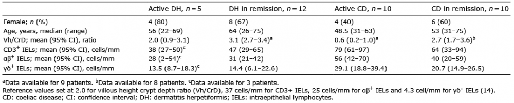
For morphological studies, one small-intestinal biopsy sample was fixed in formalin and processed for haematoxylin and eosin staining. The villous height-crypt depth ratios (Vh/CrD) were determined as described previously (32) and a ratio of ≥ 2 was considered normal. One small-intestinal biopsy was freshly embedded in optimal cutting temperature compound (OCT; Tissue-Tek, Sakura Finetek Europe, Holland), snap-frozen in liquid nitrogen and cut into 5-µm-thick sections. CD3+, αβ+ and γδ+ intraepithelial lymphocytes (IELs) in the frozen sections were detected by immunohistochemistry, as described previously. The reference values were set at 37 cells/mm for CD3+ IELs, 25 cells/mm for αβ+ IELs, and 4.3 cell/mm for γδ+ IELs (14).
Organ culture of small-intestinal mucosal biopsies was performed as described previously (33). Biopsies were cultured in RPMI-1640 medium (Invitrogen-Gibco, Paisley, UK) supplemented with 15% foetal bovine serum (Invitrogen-Gibco), 100 U/ml penicillin (Invitrogen-Gibco), 4 mM L-glutamine (Invitrogen-Gibco), 50 μg/ml insulin (Sigma-Aldrich Co, St Louis, MO, USA), 2 mg/ml glucose (Sigma-Aldrich) and 10 mM HEPES buffer (Invitrogen-Gibco) for 24 h at 37°C. From each patient one biopsy was cultured in the presence of medium only and one was subjected to a peptic-tryptic (PT) digest of gliadin (1 mg/ml), prepared, as described previously (34). Thereafter, culture supernatants were harvested and preserved at –20°C for further analysis.
IgA-class endomysial antibodies (EMA) in serum and organ culture medium were determined by an indirect immunofluoresence method using human umbilical cord as substrate (35). In the case of serum samples, a dilution of 1:≥5 was considered positive. For organ culture medium, undiluted samples were used and EMA titres were graded, according to staining intensity, as negative (–), weak positive (+), or strong positive (++).
TG2 and TG3 antibodies in serum and organ culture medium were measured by commercially available enzyme-linked immunosorbent assay kits (ELISA) (Celikey®, Phadia, Freiburg, Germany, and anti-heTG IgA ELISA, Immunodiagnostik AG, Bensheim, Germany, respectively). Measurements were performed according to the manufacturers’ instructions in serum samples diluted 1:100 and in centrifuged, undiluted organ culture supernatants. The optimal cut-off values for serum sample positivity were > 5 and > 22 AU/ml for TG2 and TG3 antibodies, respectively.
Small-intestinal mucosal IgA deposits and their co-localization with TG2 and TG3 was investigated on cryosections of small-intestinal mucosal biopsies. TG2-specific IgA deposits were stained by a direct immunofluorescence method using mouse monoclonal anti-TG2 antibody (CUB7402; NeoMarkers, Fremont, CA, USA) and fluorescein isothiocyanate (FITC)-labelled rabbit anti-human IgA antibody (Dako A/S, Glostrup, Denmark), as previously described (2). In coeliac disease, subepithelial IgA deposition can be found below the basement membrane along the villous and crypt epithelium and around mucosal blood vessels, whereas in normal small-bowel mucosa IgA is detected inside the plasma and epithelial cells (2). To determine the presence of TG3 in the small-bowel mucosa, sections were incubated with rabbit polyclonal anti-TG3 antibody (1:100; A015, Zedira, Darmstadt, Germany) for 1 h at room temperature (RT), followed by incubation with Alexa Fluor 568-conjugated goat anti-rabbit antibody (1:2000; Invitrogen) for 1 h at RT. Co-localization of TG3 with IgA was assessed by further incubating the sections with FITC-labelled rabbit anti-human IgA antibody (1:40; Dako A/S) for 15 min at RT.
Immunofluorescence staining of small-intestinal recombinant TG2- and TG3-binding cells was performed using a technique formerly established by Di Niro et al. (5) with minor modifications. Cryosections of small-intestinal biopsies 5-µm-thick were air-dried for 20 min at RT, washed with phosphate-buffered saline (PBS) and incubated with either biotinylated human recombinant TG2 (2 μg/ml; T002, Zedira) or TG3 (5 μg/ml; T024, Zedira) in 1% bovine serum albulin – phosphate-buffered saline (BSA-PBS) for 45 min RT. Biotinylation of both recombinant proteins was performed using EZ-Link® Sulfo-NHS-LC-Biotin (Thermo Scientific, Waltham, MA, USA) according to manufacturer’s instructions. After washing, rhodamine-labelled streptavidin (1:1000; KPL, Gaithersburg, MD, USA) in 10% BSA-PBS was applied for 30 min at RT. For double stainings, an additional incubation step with FITC-conjugated anti-human IgA antibody (1:40; Dako A/S) for 15 min at RT was applied.
Statistical analyses were performed using the non-parametric Wilcoxon test, Mann–Whitney test and Fisher’s test, as appropriate. Correlation was evaluated using Spearman’s correlation. A p-value < 0.05 was considered statistically significant.
Two patients with active DH reporting dietary failures had abnormal Vh/CrD representing partial villous atrophy, and the remaining patients with DH had normal villous architecture (Table I). Vh/CrD was below normal in all patients with active coeliac disease; 4 had total, 2 had subtotal, and 3 had partial villous atrophy. In contrast, all patients with coeliac disease in remission evinced normal mucosal architecture. The mean levels of CD3+, αβ+ and γδ+ T cells were above the reference level in all 4 study groups. The levels of all IEL subsets were comparable between the active DH and DH in remission groups, but lower than in both of the coeliac disease patient groups (Table I).
Serum EMA was negative in all patients with DH irrespective of disease activity. In contrast, 90% of patients with coeliac disease with active disease and 30% of patients in remission had positive serum EMA (Fig. 1A). When assessing the secretion of EMA into the organ culture medium, only one patient with active DH was observed to secrete EMA, in contrast to 80% and 20% of patients with active coeliac disease and coeliac patients in remission, respectively (Fig. 1B). TG2 antibody results in patient serum samples paralleled the EMA findings, being negative in both DH patient groups, positive in all patients with active coeliac disease and negative in patients with coeliac disease in remission (Fig. 1C). Similarly, TG2 antibody levels in the organ culture medium were either negative or very low (< 20 AU/ml) in both DH patient groups and in patients with coeliac disease in remission (Fig. 1D). In contrast, 70% of patients with active coeliac disease secreted noticeable amounts of TG2 antibodies into the organ culture medium (> 50 AU/ml). The levels of TG2 antibodies in the organ culture medium correlated with those in serum (R = 0.394, p = 0.017). PT-gliadin had no effect on the secretion of EMA or TG2 antibodies into the organ culture medium (data not shown).
Fig. 1. Levels of antibodies in the serum and organ culture medium of dermatitis herpetiformis (DH) and coeliac disease (CD) patients. (A and B) Endomysial (EMA), (C and D) transglutaminase 2 (TG2) and (E and F) TG3 antibodies (abs) in DH and coeliac disease patient serum and organ culture medium as assessed by immunofluorescence staining or enzyme-linked immunoassay (ELISA). Horizontal lines represent median values, dashed lines cut-off values for positivity provided by the ELISA kit manufacturer for serum samples. Only p-values considered statistically significant (<0.05) are indicated.
When TG2 autoantibodies were assessed in the small-intestinal mucosa, none of the patients with DH in either group was found to have TG2-bound IgA in the basement membrane or around blood vessels (Fig. 2). This was in contrast to patients with coeliac disease, of whom all patients with active coeliac disease and 60% of DH patients in remission presented with such small bowel mucosal TG2-targeting IgA deposits.
Fig. 2. Transglutaminase 2 (TG2)-targeting IgA deposits as well as recombinant TG2-binding, IgA-positive cells in the small intestine of patients with active dermatitis herpetiformis (DH) and those with coeliac disease. Small-intestinal TG2-targeting IgA deposits (A) were not present in patients with DH, but (B) were evident in patients with coeliac disease (arrow). (C) Recombinant TG2-binding IgA-positive cells were generally not detected in active patients with DH, but (D) were found in the majority of patients with active coeliac disease (arrow). TG2 or recombinant TG2 (red), IgA (green) and their co-localization (yellow) at 20× magnification. Scale bar =100 μm in all panels.
Applying an approach previously used to identify TG2-specific plasma cells in the small-intestinal mucosa of patients with coeliac disease (5, 6), we further investigated the possible presence of such IgA-positive recombinant TG2-binding cells in the small intestine of our patients with DH and those with coeliac disease. They were not found in patients with DH of either group, with the exception of one patient with DH in remission. This patient had a detectable, but low, number of recombinant TG2-binding cells. In contrast, such cells were found in 8 out of 10 patients with active coeliac disease and in 4 out of 10 patients with coeliac disease in remission (Fig. 2).
Similarly, we next addressed the autoantibody response against the other TG isoform, TG3, at serum and small-intestinal level. Serum TG3 antibody levels were below the cut-off value (> 22 AU/ml) in the majority of patients in all groups; only 1 patient with active DH and 1 with DH in remission had positive serum TG3 antibodies in addition to 3 patients with active coeliac patients and 1 in remission (Fig. 1E). Interestingly, however, TG3 antibody titres of ≥ 40 AU/ml were detected in the organ culture medium of all patients with active DH, one patient with DH in remission and one patient with active coeliac disease (Fig. 1F). There was no correlation between the TG3 antibody levels in the organ culture medium and serum (R = 0.241, p = 0.157). Moreover, PT-gliadin had no effect on the secretion of TG3 antibodies into the organ culture medium in any of the patient groups (data not shown).
When double stainings for TG3 and IgA were performed to establish the presence of IgA antibodies bound to TG3 in the small-bowel mucosa (similarly to coeliac-type TG2-bound IgA deposits), no extracellularly located TG3, and thus no TG3-targeting IgA deposits, were detected in the small-intestinal sections. Instead, we were able to detect TG3-positive cells, the majority of which were also positive for IgA (Fig. 3). A few such cells were present in 3 and 4 patients with active DH and DH patients in remission, respectively. In patients with active coeliac disease, such cells were found in 4 out of 10 patients, whereas in patients with coeliac disease in remission no such cells were detected (Fig. 3). The presence and number of the cells was not associated with TG3 antibodies in the serum or the organ culture medium.
Fig. 3. Transglutaminase 3 (TG3)-positive cells, as well as recombinant TG3-binding, IgA-positive cells in the small intestine of patients with active dermatitis herpetiformis (DH) and coeliac disease. TG3- and IgA-positive cells were present in a few patients with (A) DH (arrow), and (B) coeliac disease (arrow). Recombinant TG3-binding IgA-positive cells (arrow) were detected in (C) patients with DH, but (D) were generally absent in patients with coeliac disease. TG3 or recombinant TG3 (red), IgA (green) and their co-localization (yellow) at 20× magnification. Scale bar = 100 μm in all panels.
Finally, when the presence of IgA-positive cells binding recombinant human TG3 in the small-intestinal sections was investigated, such cells were found at low frequency in 3 out of 5 patients with active DH (Fig. 3). These 3 patients were those secreting the highest levels of TG3 antibodies into the organ culture medium (> 100 AU/ml). In addition, such cells were also detected in the one patient with active coeliac disease secreting TG3 antibodies into the organ culture medium, whereas no such cells were visualized in patients with DH in remission or the rest of the patients with coeliac disease.
The present study showed that, even though the majority of patients with active DH were negative for serum TG3 antibodies, they all secreted these antibodies into the organ culture medium. In contrast, patients with active coeliac disease had higher levels of TG3 antibodies in the serum, but low levels of secretion into the organ culture medium. Thus, the levels of TG3 autoantibodies in serum and organ culture medium did not parallel each other, in contrast to EMA and TG2 antibodies, which were mostly absent in all patients with DH, but present in patients with active coeliac disease in both type of samples.
The presence of TG2-targeting autoantibodies in the organ culture medium of coeliac disease patient-derived small-intestinal biopsies has been held to be explicable by the detachment of the autoantibodies from tissue-bound deposits and their release into the culture supernatant (28). This hypothesis seemed plausible, as all of our patients with considerable amounts of organ culture antibodies also had intestinal TG2-targeting deposits. We therefore tested whether this could also explain the presence of TG3 antibodies in the organ culture medium. We observed, however, that TG3 was not found extracellularly in the small intestine of any of our patients, which is in line with one previous study (17), and thus, the TG3 autoantibodies in the organ culture medium are not derived from detached mucosal TG3-targeted IgA deposits. Interestingly, however, a subset of patients with DH and those with coeliac disease were found to have occasional TG3-positive cells, the majority of them also being positive for IgA, but the presence and amount of these cells did not parallel TG3 autoantibodies in the culture medium. Whether these cells are antigen-presenting cells which have been suggested to express TG3 (36) remains to be determined in future studies, together with their possible involvement in DH and coeliac disease.
In addition, TG2 antibodies in the organ culture medium have been suggested to be secreted by TG2-specific plasma cells (3, 26, 37), which have recently been described in the small-intestinal mucosa of patients with coeliac disease (5, 6). In our study, those patients secreting TG2 antibodies into organ culture medium had, with the exception of one individual, recombinant TG2-binding cells in the lamina propria. Investigating the presence of TG3-antibody secreting cells in a similar manner, we detected IgA-positive recombinant TG3-binding cells in those 3 patients with DH and one patient with coeliac disease secreting the highest levels of TG3 antibodies into the organ culture medium. Even though the presence of such cells did not entirely coincide with the secretion of TG3 antibodies into the organ culture medium, our findings would indicate the TG3-binding IgA-positive cells as the source of the antibodies in the medium. It would therefore appear that, similarly to TG2-targeting antibodies (3–6), TG3 antibodies are also secreted at the small intestinal level, particularly in active DH. However, our results do not exclude the possibility that TG3 antibodies are also produced in other tissues. In fact, extraintestinal TG3 antibody secretion occurring, for instance, in bone marrow, spleen and lymph nodes could possibly explain the presence of these antibodies in the serum of those patients with coeliac disease who are negative for organ culture medium antibodies and intestinal TG3-binding IgA-positive cells.
On the other hand, the inconsistency between the general TG3 seronegativity of the patients with active DH and the presence of these antibodies in the organ culture medium along with TG3-binding intestinal cells can probably be explained by other factors. As 2 of our patients with active DH admitted regular dietary transgressions, this continued gluten intake might be sufficient to maintain antibody production locally only in the small intestine, as suggested previously (6). Moreover, it has been shown recently that, in DH, circulating TG3 antibodies can exist as immune complexes with TG3 even without free TG3 antibodies (18), which might provide another explanation for the seronegativity of the patients.
The pathognomonic IgA in DH has been suggested to be deposited in the dermis as immune complexes with TG3, which is found at this location only in DH (17). All our active DH patients and 33% of those in remission had cutaneous IgA deposits. Moreover, in active DH IgA always co-localizes with TG3 (38) and we recently showed that they disappear in parallel during a gluten-free diet, supporting their dermal deposition as immune complexes (39). Although the results of the current study point towards an intestinal origin of TG3 autoantibodies, the site of immune complex formation remains unknown.
The major strength of the present study is the well-defined cohorts of patients with DH and coeliac disease at different stages of disease. Moreover, the antibody responses against the 2 TG isoforms were studied in multiple settings, namely serum, organ culture medium and small bowel-mucosal biopsies, addressing both the presence of the antibodies as well as their possible origin. A major weakness is the particular nature of the DH patient groups. In contrast to patients with active coeliac disease, the patients with active DH were not newly diagnosed, but had active disease despite a gluten-free diet. This very likely explains many of the inconsistencies in antibody response between these patient groups, including the TG2 and TG3 seronegativity of the active DH patient group. Moreover, the patients with DH in remission had been on a gluten-free diet for a considerably longer time than the patients with coeliac disease in remission. Another limitation is the 24-h duration of the organ culture, which, even though most commonly used, may explain the unresponsiveness of autoantibody secretion to gluten in all patient groups, as also noted elsewhere (24, 25, 28, 37, 40).
Despite these limitations, our study provides important information on the autoantibody responses against TG2 and TG3 in DH and coeliac disease. Firstly, at least in our patient cohort the TG2 and TG3 autoantibody responses in serum and small-bowel mucosa were clearly different between patients with DH and those with coeliac disease. Secondly, our study revealed that, similarly to TG2 antibodies, TG3 antibodies can be detected not only in the serum, but also in the organ culture medium of small-intestinal mucosal biopsies. Most of all, our results strongly suggest that TG3 autoantibody secretion occurs in the small intestine, especially in active DH. Moreover, as the cells secreting these antibodies were found in patients with DH on a long-term gluten-free diet of up to 33 years, they must belong to an exceptionally long-lived plasma cell population (41). Even though the results of this study are not directly applicable to patients with DH at diagnosis or after short-term treatment, they provide a platform for future studies addressing this issue.
This study was supported by the Academy of Finland, the Finnish Medical Foundation, the Research Fund of the Finnish Celiac Society, the Sigrid Juselius Foundation, the Päivikki and Sakari Sohlberg Foundation, the Foundation for Pediatric Research, and the Competitive State Research Financing of the Expert Area of Tampere University Hospital.
The authors have no conflicts of interest to declare.


 Click to show fullsize
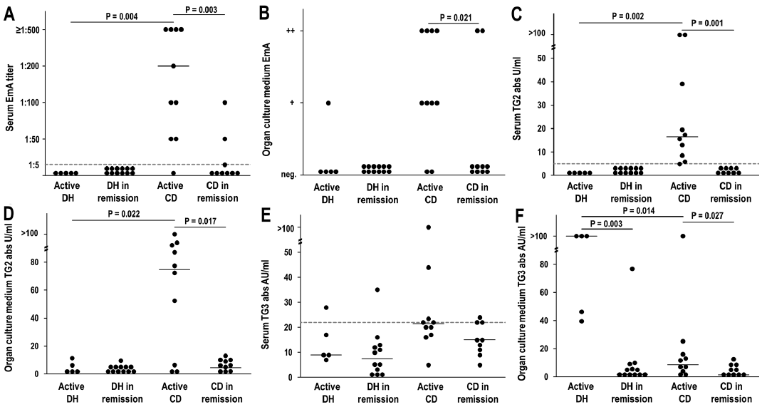 Click to show fullsize
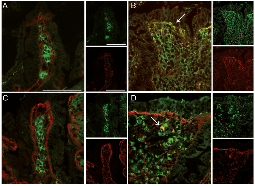 Click to show fullsize
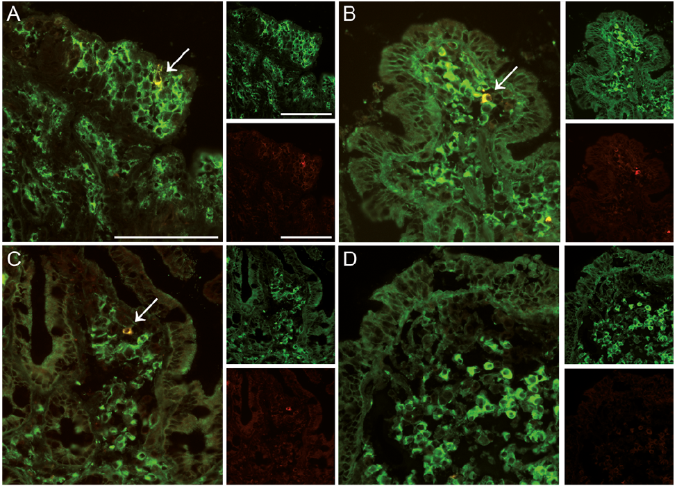 Click to show fullsize