


1Dr. Phillip Frost Department of Dermatology and Cutaneous Surgery, Miami Itch Center, University of Miami Miller School of Medicine, Miami, FL 33136, USA, and 2Department of Dermatology, Graduate School of Medical and Dental Sciences, Tokyo Medical and Dental University, Tokyo, Japan. E-mail: takiyama@miami.edu
Accepted May 29, 2019; E-published May 29, 2019
Itch, an unpleasant sensation that evokes scratching behavior, is divided into two forms: histaminergic and nonhistaminergic itch. Histaminergic itch is caused by histamine that is secreted principally by mast cells and basophils, while nonhistaminergic itch is evoked by various pruritogens other than histamine from multiple origins. Recent findings suggest that not only the interaction between pruritogens and peripheral pruriceptor terminals, but also the interplay among peripheral pruriceptor terminals, immune cells, and epidermal keratinocytes play roles in itch induction (1, 2). However, the molecular mechanisms of this interplay are not fully understood.
Signal transducer and activator of transcription (STAT)3 is a transcription factor (3) that has recently been highlighted as a key player in itch sensation. In the spinal cord, STAT3 amplifies itch sensation through promoting astrogliosis (4). While STAT3 plays critical roles in the regulation of keratinocyte function (3), whether STAT3 in the skin is also important in itch sensation is still unclear. Since keratinocytes are capable of producing itch mediators (2), keratinocyte STAT3 may also modulate itch. The aim of this study was to elucidate the involvement of keratinocyte STAT3 in itch sensation.
To examine the role of STAT3 in keratinocytes in vivo, we crossed Stat3 flox mice with K5CreERT2 mice, and tamoxifen was injected to delete STAT3 in keratinocytes. Keratinocyte-specific STAT3-depletion was confirmed through immunohistochemistry (Fig. S1). Vehicle-treated K5CreERT2:Stat3 flox/+ mice were used as controls. For further information on materials and methods, see Appendix S1. All animal experiments were approved by the Institutional Committee of the University of Miami.
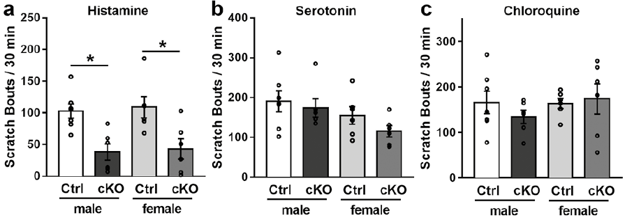
We initially assessed the in vivo effect of keratinocyte-specific STAT3 depletion on acute itch in mice. The acute itch was evoked by intradermally injecting either histaminergic or non-histaminergic pruritogen (histamine, serotonin, or chloroquine). All 3 pruritogens evoked scratching responses in control mice. Histamine-evoked scratching behavior was significantly attenuated in STAT3 conditional knockout (cKO) mice while serotonin- and chloroquine-induced scratching responses did not change (Fig. 1). We also examined the impact of keratinocyte STAT3 on chronic itch using ovalbumin (OVA)-induced atopic dermatitis (AD) murine model, which has been shown to involve nonhistaminergic itch (5). We did not find any significant reduction of OVA-induced scratching responses in STAT3 cKO mice (Fig. S2). These results demonstrate that keratinocyte STAT3 contributes to histaminergic itch, but not nonhistaminergic itch.
Fig. 1. Effects of in vivo depletion of keratinocyte STAT3 on scratching behaviors. (a) K5CreERT2:Stat3 flox/+ mice were injected with either tamoxifen (cKO) or vehicle (Ctrl). cKO mice showed attenuated histamine-induced scratching behavior in comparison with Ctrl mice. (b,c) Scratching bouts in response to serotonin (b) and chloroquine (c) did not change in cKO mice compared with Ctrl mice. Vertical bars indicate mean + SEM (n = 6–7 mice/group). * p < 0.05 versus Ctrl mice, two-tailed unpaired t-test.
To determine whether pruritogens elevate intracellular calcium in keratinocytes, we performed calcium imaging experiments on keratinocytes. In keratinocytes from control mice, intracellular calcium gradually increased and reached a maximum level 90-150 s after application of histamine and serotonin (Fig. S3a–c). However, chloroquine did not increase intracellular calcium (Fig. S3d), which is consistent with a previous report (1). The chloroquine receptor, Mas-related G protein coupled-receptor member A3, is expressed exclusively by sensory neurons, but not keratinocytes, suggesting that chloroquine evokes itch without affecting keratinocytes.
We next examined whether STAT3 is involved in intracellular calcium elevations by pruritogens in keratinocytes. The increased responses of keratinocytes to histamine and serotonin were significantly reduced in STAT3 cKO mice (Fig. S3a–c). This result was confirmed using a STAT3 inhibitor, STA-21. Keratinocytes treated with STA-21 for one hour showed a significant reduction of intracellular calcium responses compared to vehicle-treated keratinocytes (Fig. S3e, f).
A recent report has shown that histamine-induced Ca2+ influx in keratinocytes is dependent on transient receptor potential cation channel subfamily V member 4 (TRPV4), a highly calcium selective ion channel, and TRPV4-mediated Ca2+ influx is required for histaminergic itch (2). Thus, we examined the effects of a selective TRPV4 inhibitor, GSK205, on the intracellular calcium responses to histamine and serotonin in keratinocytes. The magnitude of intracellular calcium responses of keratinocytes from naive mice was significantly reduced by the application of GSK205 (Fig. S3e, f).
To investigate whether STAT3 is involved in TRPV4 expression, we performed immunostaining experiments on epidermal sheets. In epidermal sheets from control mice, TRPV4 was detected on the cell surface of keratinocytes (Fig. S4a). This signal was considerably weaker in keratinocytes of STAT3 cKO mice, while TRPV4 mRNA expressions were detected in the epidermal sheets from control mice as well as STAT3 cKO mice (Fig. S4b).
In this work, we showed the importance of keratinocyte STAT3 in histaminergic itch. We also detected that serotonin- and chloroquine-induced itch were not significantly affected by in vivo depletion of keratinocyte STAT3. These two types of itch are also implicated in chronic itch, and this result seems to be compatible with the data that scratching behavior of OVA-induced AD was not attenuated in STAT3 cKO mice.
It is somewhat surprising that depletion of keratinocyte STAT3 significantly attenuated serotonin-evoked calcium responses of the keratinocytes, but not serotonin-evoked scratching behavior. A possible explanation for these results may be that intracellular calcium increases in keratinocytes and trigger the release of the itch enhancers that can modulate itch evoked by histamine, but not serotonin. Histaminergic itch is mediated through TRPV1 in sensory neurons, whereas serotonin-induced itch is independent of TRPV1 (6). It can, therefore, be assumed that TRPV1 sensitizers (e.g., nerve growth factor, IL-6, and granulocyte-macrophage colony-stimulating factor; (7–9)) enhance histaminergic itch without affecting serotonin-induced itch. Consistent with this idea, histamine is reported to release all above-mentioned chemical messengers from keratinocytes (10–12).
Another possible explanation for the mismatch of reduced calcium signaling/unchanged scratch behavior for serotonin may be that chemical mediators produced from keratinocytes are different between histamine and serotonin. This hypothesis is supported by the finding that histamine and serotonin differently affect on keratinocytes in epidermal barrier recovery (13). Keratinocyte may release itch mediators by histamine, but not serotonin.
The intracellular calcium responses to histamine and serotonin were reduced in both keratinocytes from STAT3 cKO mice and in keratinocytes treated with the STAT3 inhibitor as well as the TRPV4 antagonist. While TRPV4 mRNA expressions were detected in the epidermal sheets from STAT3 cKO mice, TRPV4 expression was faint in keratinocytes from STAT3 cKO mice. These results suggest that STAT3 might be involved in the trafficking of TRPV4 to the plasma membrane. TRPV4-containing vesicles derived from mitochondria fuse to the plasma membrane within a few minutes to ensure constant turnover of TRPV4 (14). STAT3 may reduce TRPV4 expression by disturbing this exocytosis process. In accordance with this idea, mitochondrial STAT3 has a critical role in exocytosis in mast cells (15).
Although keratinocytes express a variety of itch mediators, how keratinocytes contribute to itch is still unclear. This study has shown that keratinocyte STAT3 play a major role in histaminergic itch, but not nonhistaminergic itch. A further study could assess the role of other signaling pathway molecules in itch via keratinocytes.
This work was supported by Pfizer grant (W1203521: GY and TA) and National Institutes of Health grant (AR063228: TA). TH has received scholarships from the Uehara Memorial Foundation, the Japanese Society of Allergology.
Conflicts of interest: GY has served in advisory board and consultant for Pfizer, Novartis, Eli Lilly, Sanofi, Galderma, Trevi, Menlo, Sienna, UCB.


 Click to show fullsize