


1Department of Dermatology and 3Department of Pathology, Paul-Sabatier Toulouse III University; IUCT-oncopole and CHU de Toulouse, 24 chemin de Pouvourville TSA 30030, FR-31059 Toulouse, 2Department of Dermatology, IUCT-oncopole, and 4Inserm UMR 1037-CRCT, Toulouse, France. E-mail: meyer.n@chu-toulouse.fr
Accepted Aug 22, 2019; E-published Aug 22, 2019
The anti-tumour efficacy of anti-programmed cell death (anti-PD1) therapy has been demonstrated in a large range of malignant neoplasms, including metastatic melanoma, advanced cutaneous squamous cell carcinoma and lung carcinoma (1). Despite presenting a favour-able safety profile, monoclonal antibodies targeting PD-1 are associated with dermatological toxicities, which affect approximately > 30% of treated patients (2). These predominantly manifest as eczema-like maculopapular rashes, lichenoid reactions, vitiligo-like lesions, or flares of psoriasis. These cutaneous adverse events generally have an immune aetiology and can, in most cases, be managed and reversed (3, 4). We report here a case of a patient developing eruptive keratoacanthoma (K-A)-like lesions induced by treatment with pembrolizumab, an anti-PD1 monoclonal antibody. Such eruptions represent an unusual cutaneous toxicity, which has, until now, been very rarely reported.
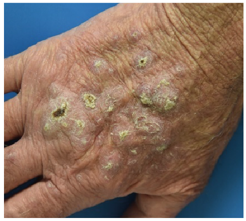
An 85-year-old man was treated with pembrolizumab for metastatic bronchial squamous cell carcinoma (SCC). After the second treatment cycle he developed hyperkeratotic nodular lesions (more than 30 lesions in total) on the dorsal region of his hands, knees and legs (Fig. 1). The mucous membranes and scalp were not affected. The patient had no prior history of skin disease. However, he had experienced chronic exposure to ultraviolet (UV) light during outdoor leisure activities. Histopathological analysis of 3 eruptive lesions showed a similar pattern of infiltrative well-differentiated SCCs. Immunostaining identified a predominantly CD3+/CD4+ associated T-lymphocyte infiltrate of the basement membrane, with no lichenoid involvement. Anti-PD1 staining was weakly positive. After clinical-pathological concertation, the lesions were diagnosed as keratoacanthoma-like lesions. Since an objective regression of the lung cancer was observed, treatment with pembrolizumab was maintained. Clobetasol cream (once per day) was prescribed for topical application to the dorsal region of the hands, knees and legs, and the lesions gradually regressed over a few weeks. Complete regression, without scarring, was noted after 3 months (Fig. 2). At the 6-month follow-up, no recurrence or progression of skin lesions was observed.
Fig. 1. Keratosis eruption after 2 cycles of immunotherapy with pembrolizumab.
Fig. 2. Clearance of lesion with propionate clobetasol.
We report here an additional case of eruptive keratoacanthoma-like eruption induced by pembrolizumab. Anti-PD1 antibodies play a major role in the management of melanoma and locally-advanced or metastatic cutaneous SCCs. The paradoxical development of eruptive keratoacanthoma-like lesions has been reported recently in 7 patients treated with pembrolizumab, and in 5 patients treated with nivolumab (5–10). Similar to the present case, eruptive keratoacanthoma-like lesions induced by immunotherapy are reported to appear early, between 2 and 18 months after the first infusion. In previously reported cases, the median age of patients was comparable (80 years old) and clinical eruption also presented with multiple violaceous, hyperkeratotic nodules, papules and plaques on the UV-exposed extremities. In the same way, pathology analysis of lesions showed atypical keratinocytes, immune infiltrates composed of CD3+ T cells with scattered CD20 B cells. As observed in our patient, lymphocytes were weakly positive on PD-1 immunostaining. In general, patients were treated with topical clobetasol cream once per day. In addition, 3 patients were also treated with superficial cryotherapy, one patient with 5-fluorouracil intralesional injections, one patient with intralesional steroids, 3 patients with curettage and two patients were not given any treatment. The lesions disappeared in a few months. Similarly to previous case reports, we also initiated treatment with topical clobetasol alone, which resulted in significant regression of the K-A-like lesions.
One may hypothesize that anti-PD1 raises an immunological response against sub-clinical epidermal UV-induced dysplasia, resulting in transient epithelial proliferation. This hypothesis is strengthened by the age of patients with more predispositions to cutaneous dysplasia and the location of lesions on UV-exposed areas of the skin. Moreover, we speculate that the mechanism driving the development of secondary skin neoplasms is similar to those observed in tumour pseudo-progression associated with immune checkpoint inhibitors (11). This hypothesis is supported by the fact that, in all patients, the lesions improved with topical steroid therapy, which is not the usual treatment for keratoacanthomas. In addition, pembrolizumab effectively treated the bronchial SCC in our patient. Indeed, cross-reactivity of T cells with tumour epitopes and dysplastic epidermal cells may have participated in the induction of an effective immune reaction in UV-exposed skin, which clinically translated into eruptive keratoacanthomas-like eruption. Finally, pembrolizumab has demonstrated efficacy in the treatment of cutaneous SCCs and the long-lasting clearance of lesions without requiring any additional specific therapeutic procedures, which strongly suggests that pembrolizumab participated in the treatment of the skin lesions. Topical corticosteroids are indicated to control skin inflammatory symptoms. The anti-tumour activity of pembrolizumab may control tumour proliferation, whereas the topical corticosteroids can control the temporary cutaneous inflammation (like oral corticosteroids in the case of other side-effects) (12).
Conclusion
The paradoxical eruption of multiple keratoacanthoma-like lesions in UV-exposed areas of the skin seems to be induced by anti-PD-1 therapy. It may be considered as a pseudo-progression of subclinical high-grade intra-epidermal dysplasia. This case may point to the presence of a rare cutaneous side-effect of anti-PD1 receptor antibody therapy that generally resolves spontaneously, allowing for anti-PD-1 treatment to be maintained and the skin lesions to be treated with minimally invasive therapeutic approach.


 Click to show fullsize
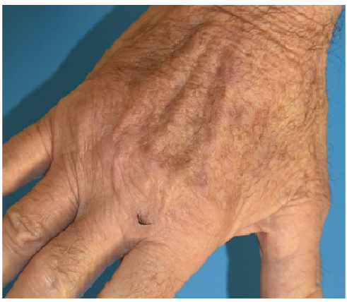 Click to show fullsize