


1Department of Dermatology, The University of Tokyo Graduate School of Medicine, Tokyo, 2Department of Dermatology, St Marianna University School of Medicine, Kanagawa, and 3Department of Dermatology, International University of Health and Welfare, Chiba, Japan
In many malignancies, dysregulation of the Notch path-ways, composed of 4 Notch receptors (Notch1–4) and 5 Notch ligands (Jagged1–2, Delta-like ligand-1, 3–4), is associated with their development. In mycosis fungoides, interaction between Notch1 and Jagged1 is known to activate the Notch pathways and promote the proliferation of tumour cells. However, the involve-ment of other Notch ligands has not been reported. This study investigated the roles of Delta-like ligand 4 in mycosis fungoides. Delta-like ligand 4 mRNA levels in lesional skin of patients with mycosis fungoides were significantly elevated compared with those of normal controls, and correlated with disease-specific mortality. Immunohistochemical staining demonstrated prominent expression of Delta-like ligand 4 on vascular endothelial cells and tumour cells in mycosis fungoides lesional skin. In addition, Delta-like ligand 4 augmented the proliferation of cutaneous T-cell lymphoma cell lines. These results suggest that enhanced Delta-like ligand 4 expression may contribute directly to the progression of mycosis fungoides through proliferating tumour cells.
Key words: delta-like ligand 4; mycosis fungoides; cutaneous T-cell lymphoma; Notch homolog 1.
Accepted Jan 9, 2020; Epub ahead of print Jan 13, 2020
Acta Derm Venereol 2020; 100: XX–XX.
Corr: Tomomitsu Miyagaki, Department of Dermatology, St Marianna University School of Medicine, 2-16-1, Sugao, Miyamae-ku, Kawasaki-shi, Kanagawa 216-8511, Japan. E-mail: asahikari1979@gmail.com
In many malignancies, including mycosis fungoides, dysregulation of the Notch pathways has been reported to be associated with their development. Thus, Notch-specific monoclonal antibodies against Notch1, Notch2 or Notch3 have been developed for the treatment of malignancies. This study found that expression levels of Delta-like ligand 4, one of the Notch ligands, was increased in lesional skin of patients with mycosis fungoides, and that Delta-like ligand 4 had the capacity to induce proliferation of cutaneous T-cell lymphoma cell lines. These results suggest that not only monoclonal antibodies against Notch receptors, but also anti-Delta-like ligand 4 antibodies, may be a useful therapeutic tool for mycosis fungoides.
Mycosis fungoides (MF) is the most common type of cutaneous T-cell lymphoma (CTCL) (1). MF is characterized by malignant proliferation of CD4+ T cells with epidermotropism in the skin and has a typically prolonged clinical course. In limited cases, progression over years through patch, plaque and tumour stages, followed by lymph node and visceral involvement, is observed (2, 3). The management of such advanced MF is challenging due to lack of effective therapies.
The Notch pathways with 4 Notch receptors (Notch1–4) and 5 Notch ligands (Jagged1–2, Delta-like ligand (DLL)-1, 3–4) play critical roles in cell development, differentiation, proliferation, and death through activating target genes, including MYC, nuclear factor-κB, cyclin D1, and BCL2 (4, 5). In many haematopoietic and solid malignancies, dysregulation of the Notch pathways has been observed. The dysregulated pathways can be oncogenic in most malignancies, while in some malignancies, such as B-cell acute lymphoblastic leukaemia, cutaneous squamous cell carcinoma, and small cell lung cancer (1), Notch can function as a tumour suppressor. As for T-cell malignancies, T-cell acute lymphoblastic leukaemia has been widely studied and it is evident that crosstalk between DLL4 and Notch1 or Notch3 has oncogenic roles (6–8). In MF and Sézary syndrome, a leukaemic variant of CTCL, prominent Notch1 expression on tumour cells, especially in more advanced stages, is confirmed by immunohistochemistry (9). In addition, specific down-regulation of Notch1, but not Notch2 and Notch3, induced apoptosis in the cell line derived from Sézary syndrome (9), suggesting that the Notch1 pathway is associated with CTCL development. More recently, Gallardo et al. have revealed that Jagged1 overexpression due to the methylation of Notch-related microRNA-200c is involved in Notch1 pathway activation in tumour stage MF (10). However, the expression and function of other Notch ligands in MF have not been reported. This study examined the involvement of DLL4, which is important for progression of another T-cell malignancy, T-cell acute lymphoblastic leukaemia, in MF development.
Patients
All patients with MF enrolled in this study were given diagnoses according to the current International Society of Cutaneous Lym-phoma/European Organisation for Research and Treatment of Cancer ((ISCL/EORTC) criteria (1, 11). Skin samples for messenger RNA (mRNA) extraction were obtained from lesional skin of patients with MF (n = 52: stage IA 13, stage IB 8, stage IIA 3, stage IIB 18, stage IIIA 8, and stage IVB 2; mean ± standard deviation (SD) age: 57.8 ± 14.8 years; 41 males and 11 females), and healthy controls (n = 14; 49.0 ± 14.9 years; 6 males and 8 females). Skin samples for immunohistochemistry were collected from lesional skins of 22 patients with MF (stage IA 4, stage IB 6, stage IIB 6, stage IIIA 5, and stage IVA2 1; 52.5 ± 13.5 years; 17 males and 5 females), and 8 healthy controls (24.9 ± 11.2 years; 2 males and 6 females). Serum samples were obtained from 32 patients with MF (stage IA 11, stage IB 1, stage IIA 2, stage IIB 10, stage IIIA 5, stage IVA1 1, and stage IVA2 2; 58.6 ± 15.5 years; 22 males and 10 females), and 18 healthy control subjects (50.3 ± 13.6 years; 7 males and 11 females). Patients were subgrouped into early stage (IA, IB, and IIA) and advanced stage (IIB, IIIA, IIIB, IVA1, IVA2, and IVB) according to disease staging with TNMB classification. The healthy controls had no history of allergy, atopic dermatitis, psoriasis, or CTCL. All samples were collected at diagnosis during daily clinical practice. The medical ethics committee of the University of Tokyo approved all described studies and the study was conducted according to the principles of the Declaration of Helsinki. Written informed consent was obtained to use blood and skin samples from patients and healthy controls.
Real-time quantitative RT-PCR assay
Total messenger RNA (mRNA) was obtained from human skin samples with RNeasy Fibrous Tissue Mini Kit (QIAGEN, Valencia, CA, USA). Complementary DNA was synthesized using ReverTra Ace® qPCR RT Master Mix (TOYOBO, Osaka, Japan). mRNA levels were analysed using real-time quantitative RT-PCR method with THUNDERBIRD SYBR qPCR Mix (TOYOBO) on an ABI Prism 7000 sequence detector (Applied Biosystems, Foster City, CA, USA) according to the manufacturers’ instructions. All samples were analysed in parallel for glyceraldehyde-3-phosphate dehydrogenase (GAPDH) gene expression as an internal control. The relative expression levels of each gene were determined by the 2–ΔΔCT method. Primers for human DLL4 and GAPDH were as follows: DLL4 forward, 5’-ACA ACT TGT CGG ACT TCC AG-3’ and reverse, 5’-CAG CTC CTT CTT CTG GTT TG-3’; GAPDH forward, 5’-ACC CAC TCC TCC ACC TTT GA -3’ and reverse, 5’-CAT ACC AGG AAA TGA GCT TGA CAA-3’. All samples were analysed in parallel for GAPDH gene expression as an internal control. The relative expression levels of each gene were determined by the 2-ΔΔCT method.
Immunohistochemistry
Briefly, 5-μm thick tissue sections from formaldehyde-fixed and paraffin-embedded samples were dewaxed and rehydrated. These sections were then stained with rabbit anti-human DLL4 (1:100, Abcam, Cat #ab176876), anti-thymocyte selection-associated high mobility group box factor (TOX; 1:200, ATLAS ANTIBODIES, Cat # HPA018322), or isotype-matched control antibody, followed by ABC staining (Vector Lab, Burlingame, CA, USA). Diaminobenzidine was used for visualizing the staining, according to the manufacturer’s instructions. The positivity of vascular staining was ranked as 0–3 (0, absent; 1, weak; 2, moderate; 3, strong).
Enzyme-linked immunosorbent assay (ELISA)
DLL4 in sera was quantified using DLL4 ELISA kit (Cloud-Clone Corp., Katy, TX, USA). These assays employ the quantitative sandwich enzyme immunoassay technique. Optical densities were measured at 450 nm using a Bio-Rad Model 550 microplate reader (Bio-Rad Laboratories, Hercules, CA, USA). The concentrations were calculated from the standard curve generated by a curve-fitting program according to the manufacturer’s instructions.
In vitro experiments
MyLa (mycosis fungoides cell line), HH (aggressive cutaneous T-cell lymphoma cell line) and Hut78 (Sézary syndrome cell line) cells were kindly provided by Dr Kazuyasu Fujii (Department of Dermatology, Kagoshima University, Kagoshima, Japan). The cells were cultured in RPMI 1640 containing 10% foetal bovine serum and supplements (penicillin G sodium, streptomycin sulphate and amphotericin B). CTCL cell lines were plated onto 96-well plates at 4×10⁴ cells/200 μl per well. The cells were stimulated with recombinant DLL4 (1 or 5 μg/ml; R&D Systems), and incubated at 37°C and 5% CO2 for 24 or 48 h. Viable cells were counted by trypan blue exclusion.
Statistical analysis
Statistical analysis was performed using Prism Version 7 software (GraphPad, San Diego, CA, USA). Statistical analysis was carried out with the Mann–Whitney U test for comparison of 2 groups. Correlation coefficients were determined using the Spearman’s rank correlation test. In the survival analysis, the median value was set as the cut-off for each factor. The survival rates were calculated using the Kaplan–Meier method, and the rates of the 2 groups were compared using a log-rank test. Multivariable Cox proportional hazards model was used to assess the association between DLL4 mRNA levels in lesional skin and patient survival. p-values of < 0.05 were considered statistically significant.
Delta-like ligand 4 mRNA expression in lesional skin of mycosis fungoides
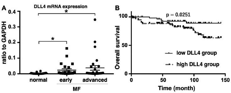
To assess DLL4 involvement in MF development, the study first examined mRNA expression levels of DLL4 in lesional skin of MF and normal skin. DLL4 mRNA levels were significantly elevated both in early MF skin and advanced MF skin compared with normal skin, while there was no significant difference in the levels between early MF skin and advanced MF skin (Fig. 1A). The study subsequently examined whether DLL4 mRNA levels correlated with patient survival. Kaplan–Meier curves with a cut-off point of 0.0030, which was the median value of MF patients, are shown in Fig. 1B. As shown in Fig. 1B, patients with high DLL4 mRNA levels (> 0.0030) had a significantly lower survival rate than those with low DLL4 mRNA levels (0.0030 and less) assessed by log-rank test (hazard ratio 0.333; 95% confidence interval (95% CI) 0.139–0.801; p = 0.025). To more precisely examine the association between DLL4 mRNA levels and patient survival, multivariate analysis was performed including factors that were reported to be associated with prognosis in patients with MF, as shown in Table I (12–14). In addition to older age, advanced stage, and presence of large cell transformation, high DLL4 mRNA levels were associated with a poor prognosis (Table I). Thus, DLL4 mRNA expression levels were upregulated in lesional skin of patients with MF and correlated with disease-specific mortality, suggesting that DLL4 is associated with development of MF.
Fig. 1. Delta-like ligand 4 (DLL4) mRNA expression in lesional skin of mycosis fungoides (MF) and normal skin. (A) DLL4 mRNA expression levels in lesional skin of early MF, advanced MF and normal skin. *p < 0.05. (B) Kaplan–Meier analysis for disease-specific survival of patients with MF with high levels of DLL4 mRNA (> 0.0030; n = 26) and those with low levels of DLL4 mRNA (≤ 0.0030; n = 26).
Table I. Multivariate analysis
Immunohistochemical staining of Delta-like ligand 4 in lesional skin of patients with mycosis fungoides
Samples of MF lesional skin were immunolabelled for DLL4. Immunohistochemical staining revealed that vascular endothelial cells expressed DLL4 in both normal skin and MF skin (Fig. 2A, F, G). The intensity of vessel staining of DLL4 was more obvious in advanced MF (Fig. 2H). In addition, it was found that epidermotropic cells and dermal infiltrating cells with atypical nuclei were positive for DLL4 (Fig. 2B, C). To examine whether such DLL4-positive cells were tumour cells, the serial sections were then stained with the antibody to TOX, which was reported to be a useful diagnostic marker in MF (15, 16). It was found that DLL4-positive cells and TOX-positive cells showed similar distribution (Fig. 2B–E), suggesting that MF tumour cells expressed DLL4. Thus, endothelial cells and tumour cells were the main sources of DLL4 in MF skin.
Fig. 2. Immunohistochemical staining for Delta-like ligand 4 (DLL4) in lesional skin of patients with mycosis fungoides (MF) and normal skin. (A–C) Immunohistochemistry for DLL4 in lesional skin of patients with MF. (D, E) Immunohistochemistry for TOX in lesional skin of patients with MF. (F) Immunohistochemistry for DLL4 in normal skin. Representative figures from 22 patients with MF and 8 healthy controls, respectively. (G) Representative control staining using isotype-matched antibody. (H) Immunohistochemical scoring of DLL4-positive endothelial cells. *p < 0.05, **p < 0.01. (Magnification: A, F, G: x200, B–E: x400).
Serum Delta-like ligand 4 levels in patients with mycosis fungoides
This study also measured serum DLL4 levels in patients with MF and healthy controls. Unfortunately, serum DLL4 levels in patients with MF were comparable to those of healthy controls (Fig. 3A). In addition, there were no significant differences in serum DLL4 levels between healthy controls and patients with MF even when subgrouped into early and advanced stages (Fig. 3B). Thus, serum DLL4 levels in patients with MF showed no significant change compared with healthy controls, suggesting that DLL4 may act mainly in lesional skin in patients with MF.
Fig. 3. Serum Delta-like ligand 4 (DLL4) levels in patients with mycosis fungoides (MF) and healthy controls. (A) Serum DLL4 levels in patients with MF and healthy controls. (B) Serum DLL4 levels in patients with early MF, advanced MF, and healthy controls.
Delta-like ligand 4 augmented the proliferation of cutaneous T-cell lymphoma cell lines
To assess the direct effect of DLL4 on proliferation of CTCL cells, this study finally stimulated CTCL cell lines with DLL4. As expected, DLL4 promoted the proliferation of MyLa, HH and Hut78 cells, all of which are reported to express Notch1 (9, 10), in a dose-dependent manner (Fig. 4). Thus, DLL4 can be involved in MF development through proliferating tumour cells.
Fig. 4. Delta-like ligand 4 (DLL4)-induced proliferation of cutaneous T-cell lymphoma (CTCL) cell lines. CTCL cell lines were cultured with or without DLL4 and cell count was performed after 24 or 48 h. The data are presented as the mean ± standard error of the mean in each group. *p < 0.05, **p < 0.01.
This study revealed that DLL4 mRNA expression is significantly upregulated in MF lesional skin and that tumour cells and endothelial cells express DLL4 (Figs 1 and 2). Similarly, augmented DLL4 expression in endothelial cells and tumour cells has been reported in various types of malignancies, such as colon cancer, ovarian cancer, and pancreatic ductal carcinoma (17–19). In addition, a recent meta-analysis revealed that DLL4 overexpression implies poor survival in pancreatic ductal carcinoma (20). Likewise, a significant correlation between high DLL4 mRNA expression in lesional skin and increased disease-related mortality was found in patients with MF (Fig. 1). Considering that Notch1 expression is detected in MF tumour cells (9, 10), the results of the current study suggest that high DLL4 expression in dermal endothelial cells and tumour cells may be associated with MF development through activating Notch signalling in tumour cells. Indeed, this study found that exogenous DLL4 administration promoted the proliferation of CTCL cell lines in a dose-dependent manner (Fig. 4).
As Notch signalling pathways are deeply connected to proliferation, metastasis and drug resistance in many malignancies (4, 5), Notch-specific monoclonal antibodies against Notch1, Notch2, or Notch3 have been developed (21). Moreover, anti-Notch ligand antibodies are also currently in development. Among them, anti-DLL4 antibodies are the most studied. Multiple early-stage clinical trials of demcizumab, enoticumab, and MedI0639, both of which are anti-DLL4 monoclonal antibodies, for various cancers have been designed and implemented (4, 22–25). Our results indicate that, in addition to antibodies against Notch1, which has been regarded as a potential therapeutic target in MF since before (9), such anti-DLL4 antibodies can also be useful therapeutic agents for MF.
In summary, this study showed that expression of DLL4 was upregulated in lesional skin of patients with MF and that DLL4 promoted the proliferation of CTCL cell lines. These results suggest that DLL4 may be directly associated with development of MF through proliferating tumour cells and might be a therapeutic target in MF.
The authors thank Tamami Kaga for technical assistance. This work was supported in part by grants from the Ministry of Education, Culture, Sports, Science and Technology in Japan (16K19709).
The authors have no conflicts of interest to declare.


 Click to show fullsize
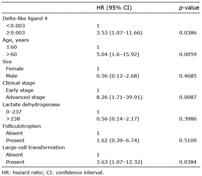 Click to show fullsize
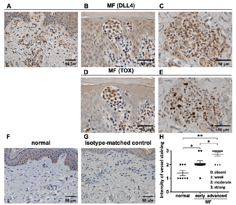 Click to show fullsize
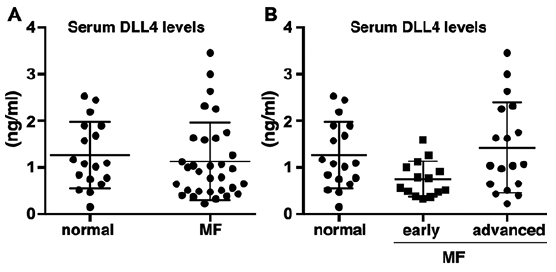 Click to show fullsize
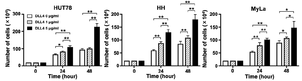 Click to show fullsize