


1Department of Dermatology and Allergy, and 2Institute of Pathology, Ludwig Maximilian University (LMU), DE-80337 Munich, Germany. *E-mail: markus.reinholz@med.uni-muenchen.de
Accepted Jan 9, 2020; Epub ahead of print Jan 17, 2020
Acta Derm Venereol 2020; 100: adv00061
More than 40 out of 200 identifiable human papillomavirus (HPV) types specifically infect the epithelia of the anogenital tract (1). Consequently, these HPV infections can manifest as condylomata acuminata (CA), also known as genital warts, which usually appear as flesh-coloured, verrucous papules or plaques in the perianal area (2). HPV types are divided into “low risk” (lr) and “high risk” (hr, oncogenic) types, based on the associated risk of neoplastic transformation (3). Although it is the most common sexually transmitted disease worldwide (4), recent studies suggest that the majority of affected children acquire HPV infection from non-sexual contact (5, 6). Therefore, the debate about HPV infection in children is moving away from the mode of transmission, and is concentrating more on the prevalence of HPV (7). Epidemiological data on CA in children are difficult to assess, and the prevalence of this condition in juvenile patients remains unknown (2). The main goals of this retrospective study were to define the epidemiological characteristics and the HPV genotype distribution in a group of juvenile patients presenting with CA in our university hospital.
Following ethical approval (UE No. 17-167) granted by the Ethics Committee of Ludwig-Maximilian-University (LMU), Munich, Germany, patient data were collected from the pathology database of LMU. A total of 17 patients, all 18 years of age or younger, presenting with histologically confirmed CA between 2003 and 2017, were retrospectively included for further analysis. Tissue samples of 13 matching patients with histologically normal anogenital skin were examined as controls. Differentiation according to sex, age and localization of the sample collection, was performed. DNA was extracted from formalin-fixed paraffin-embedded tissue in the form of 10-μm thick unstained slides. Amplification of HPV-DNA was performed by PCR using 2 pairs of primers obtained from the LCD-Array HPV Type 3.5 kit (Chipron GmbH, Berlin, Germany). Amplified HPV-DNA was used for hybridization with the LCD-Array HPV Type 3.5 kit detecting 32 α-HPV types (6, 11, 16, 18, 31, 33, 35, 39, 42, 44, 45, 51, 52, 53, 54, 56, 58, 59, 61, 62, 66, 67, 68, 70, 72, 73, 81, 82, 83, 84, 90, 91) according to the manufacturer’s instructions, evaluated using the company’s SlideReader V12 software (Chipron GmbH, Berlin Germany).
Out of the 17 patients with CA, 4 were males (23.5%) and 13 females (76.5%), age range 2–18 years, mean age of all patients 12.6 years. All male patients presented with CA in the perianal region, while the penile shaft was not affected. The youngest boy was 8 years old and the oldest 17 years old. On the other hand, the manifestations of CA in female patients were more variable, with the most common being in perianal (38.5%), cervical (23.1%), vulvar (23.1%) and vaginal (15.4%) areas. In addition, in the patient group under the age of 14 years, 6 out of 7 (85.7%) children presented with genital warts in the perianal area. The predominantly affected anatomical regions in the patient group aged 14 years or older were cervix, vulva and vagina (70.0%) (Table SI).
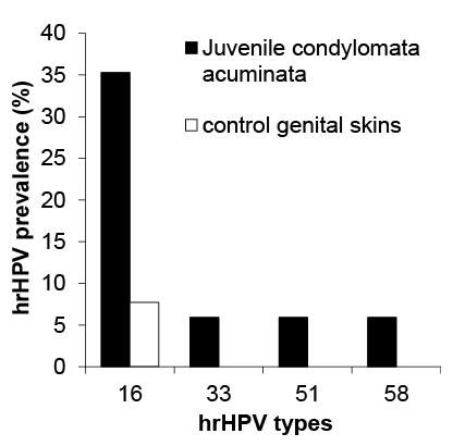
Overall, HPV-DNA was detected in all 17 patients presenting with CA. Slightly more than half of these patients (52.9%) were infected with hrHPV types in addition to the lrHPVs. The most commonly identified hrHPV type was HPV 16, which was present as a coinfection in 6 patients (35.3%) with CA, and in only one healthy patient (7.7%) as a single infection (Fig. 1). In 7 (53.8%) healthy control samples no HPV-DNA could be detected. LrHPVs could be identified in healthy controls, which, however, remained at a much lower rate (38.5%) compared with the CA skin samples (100.0%). HPV 6 was by far the most commonly detected type (94.1%), followed by HPV 11 (23.5%), in children with CA (Table I).
Fig. 1. Prevalence of high-risk human papilloma-virus (hrHPV) genotypes in juvenile condylomata acuminata (CA) vs. control genital skin samples. Vertical bars indicate the percentage of hrHPV genotypes in juvenile CA (n = 17) and control genital skin samples (n = 13).
Table I. Frequency distribution of human papillomavirus genotypes in juvenile condylomata acuminata vs. control genital skin samples
This study revealed hrHPV coinfection in more than half of the juvenile patients with CA (52.9%) and in only one (7.7%) control patient. Notwithstanding the increase in reporting, data from paediatric anogenital samples in the literature remain sparse and often contradictory. This can be explained by different socio-demographic profiles of the populations studied, the limited number of cases, the sensitivity of the HPV test and the quality of the analysis itself (7, 8). A 7-year study of children under the age of 12 years reported hrHPV (type 16) detection in only 1, and lrHPVs (type 6 and 11) in 11 of the 40 patients undergoing wart excision and HPV genotyping by PCR (9). Another review of nearly 200 children with confirmed HPV-DNA in their CA lesions reported a detection rate of only 4% for HPV 16 or 18 and 56% for HPV 6 or 11 (7). In comparison with the previous reports, the rate of oncogenic HPV subtypes detected in our current study must be regarded as remarkably high. The main reasons for this could be selection bias due to the small sample cohort originating from a tertiary referral hospital, as well as the inclusion of adolescents up to the age of 18 years into our analysis, who have a higher probability of becoming sexually active. Furthermore, the modern microarray-based genotyping used in this study may have higher sensitivity and specificity for identification of a wider range of hrHPV subtypes compared with the technologies applied in the aforementioned studies.
In our analysis we were also able to assess the HPV detection rate of anogenital skin lesions of control patients without CA. However, due to the retrospective design of this study, we were unable to conduct follow-up visits to determine the natural course of the HPV infections in our patients. The current study was also limited by the relatively low number of patients in the analyses and the lack of information about underlying diseases or probable causes of infection.
Given that sexual transmission is more likely with cervical and vaginal warts (1), this transmission mode should be considered in our study for children 14 years of age or older in particular, who were predominantly affected in these anatomical regions. Previous reports have shown that other methods of transmission may account for the majority of HPV infections in paediatric patients (5, 6). Children may acquire HPV during non-sexual contact with a caregiver (heteroinoculation), from themselves (autoinoculation), perinatally or prenatally, or by transmission via fomites, such as contaminated towels or underwear, which can occur at any age (5, 6).
The mean age of children and adolescents presenting with CA ranged from 2.8 to 5.6 years in one study (2), and spontaneous regression has been reported in only half of the cases in another (10). The other half of estimated children with persistent CA would have to wait until the age of 9 years for HPV vaccination (11, 12). Interestingly, we observed a surge in the prevalence of coinfections with persisting hrHPV subtypes in patients with CA after the age of 8 years. Oncogenic HPV types, especially HPV 16, have a longer time to clearance than other subtypes (13), and these factors may directly promote HPV-associated oncogenesis in the future.
The results of our study show a high prevalence of oncogenic HPV types in juvenile CA samples. Our findings suggest that juvenile patients with CA bear the risk of a coinfection or sequential infection with hrHPV. Therefore, special follow-up methods for children with HPV-positive family members or caregivers, as well as lowering the age for HPV vaccination, should be assessed in larger studies.
The authors would like to thank Ursula Puchta and Latifa Birrou for technical assistance.
Conflicts of interest. MR received speaker’s honoraria from MSD, GSK and MEDA/Mylan. The other authors have no conflicts of interest to declare.


 Click to show fullsize
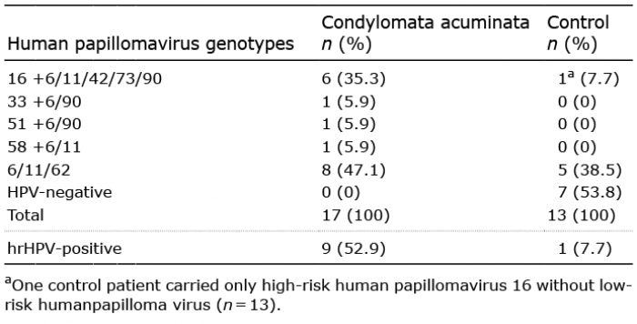 Click to show fullsize