


1Department of Dermato-Oncology/Dermatology, National Hospital Organization Kagoshima Medical Center, Shiroyama-cho, Kagoshima, Kagoshima 892-0853 and 2Department of Dermatology, Kyoto University Graduate School of Medicine, Kyoto, Japan. *E-mail: kom.takaya@gmail.com; shigeto0302@gmail.com
Accepted Oct 28, 2020; Epub ahead of print Nov 2, 2020
Acta Derm Venereol 2020; 100: adv00335.
doi: 10.2340/00015555-3681
Photosensitive dermatitis is clinically recognized as sunlight-induced dermatitis. It develops through 2 mechanisms: phototoxicity and photoallergy. Of these, photoallergic dermatitis is a type IV hypersensitive photoreaction against an external or internal antigen, which is mediated largely by ultraviolet A (UVA) (1, 2). Although various antigens, including antibiotics and non-steroidal anti-inflammatory drugs (NSAIDs) are reported to induce photosensitivity (1), there is no report that indicates the possibility of immune checkpoint inhibitor-associated photosensitivity. We report here a case of photosensitive dermatitis possibly induced by nivolumab/ipilimumab combination therapy (NIV/IPI) in a patient with malignant melanoma.
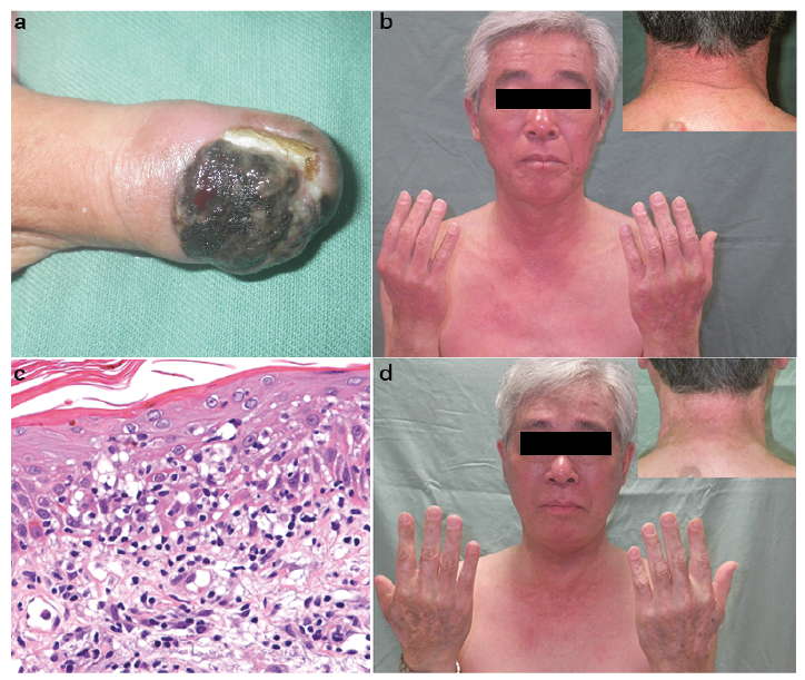
A 64-year-old, previously healthy, male developed nail apparatus melanoma of the right thumb (Fig. 1a). He underwent wide local excision and sentinel lymph node biopsy. Histological examination showed no evidence of metastasis and he was followed up carefully. After 7 months, positron-emission tomography/computed tomography revealed multiple metastases in the brain. Soon after gamma knife treatment (24 Gy/1 Fr) for brain metastases, NIV/IPI therapy was initiated.
Fig. 1. Clinical and histological images of the patient. (a) Primary lesion. (b) Worsened erythema and erosive lesion on the back of the neck. (c) Histological findings of erythema on the right forearm. Mild acanthosis, focal parakeratosis, and a few necrotic keratinocytes visible within the spongiotic epidermis. Within the superficial dermis, there is a perivascular, predominantly lymphocytic infiltrate. (d) One month after treatment with oral prednisolone, the erythema was ameliorated. Permoission is given by the patient to publish these photos.
One week after the first NIV/IPI therapy, red, palpable papules appeared on the patient’s trunk, and he was treated with topical difluprednate ointment. Immediately after the second NIV/IPI therapy, the erythema augmented and was predominantly localized in the sun-exposed area (Fig. 1b). Photo-tests showed a markedly decreased minimal erythema dose (MED) of 3 J/cm2 UVA. No obvious decreases in the MED of UVB or visible light were observed. The histological appearance was consistent with that of photosensitive dermatitis (Fig. 1c). Because the patient did not take any other medications or have any apparent episodes of complications with viral or bacterial infection before and during NIV/IPI therapy, this case was diagnosed as photosensitive dermatitis possibly induced by NIV/IPI therapy. Because erythema became erosive and spread from the distal to the proximal area, NIV/IPI therapy was discontinued and oral prednisolone treatment for photosensitive dermatitis was initiated (60 mg/day, gradually tapered to 20 mg/day over 4 months). After amelioration of the skin eruption (Fig. 1d), photo-tests were performed again, which confirmed the persistence of UVA sensitivity with a MED of 3 J/cm2. Although NIV/IPI therapy was discontinued and the patient has not taken any other anti-cancer agents over 7 months’ follow up, his melanoma has been well controlled.
In this case, the patient developed severe skin erythema during NIV/IPI therapy. From the clinical distribution of the erythema, his medication history, hypersensitivity to UVA and histopathological features (3), a diagnosis of photosensitive dermatitis was considered. Since the patient received no oral medication during NIV/IPI therapy, the possibility of hypersensitivity against external antigens with oral administration was excluded. Therefore, the patient was hypothesized to have developed photosensitivity induced by an autoimmune response to an endogenous antigen. His symptoms may be associated with NIV/IPI treatment because he had not exhibited any photosensitive symptoms before NIV/IPI therapy, although he often went outdoors for his hobby of insect collecting.
Previous reports suggest another immunomodulator has potential to induce photosensitive dermatitis (1, 4). Mogamulizumab, a monoclonal antibody targeting CCR4, was shown to induce photosensitive dermatitis (4), possibly due to serving as an immunomodulator, but not as a photoantigen. Therefore, photoallergic dermatitis may have occurred, not due to an external antigen but due to an internal antigen. This might indicate that an immunomodulator including NIV/IPI could induce immune-related photosensitive dermatitis.
In conclusion, we report here the first case of photosensitive dermatitis during NIV/IPI therapy possibly occurring as an immune-related adverse event. In contrast to previously reported photoallergic dermatitis, which is generally resolved after withdrawal of the culprit drugs (5), the finding of a decrease in the MED of UVA in photo-tests, 2 times, 4 months after the last administration of NIV/IPI, suggests that this immune-related side-effect could last longer than that of common photosensitive dermatitis.
The authors have no conflicts of interest to declare.


 Click to show fullsize