


1INSERM 1246-SPHERE, University of Tours, University of Nantes, 2Department of Neonatalogy, 3Department of Pathology, and 4Department of Dermatology, Unit of Paediatric Dermatology, FR-37044 Tours Cedex 9, France. E-mail: annabel.maruani@univ-tours.fr
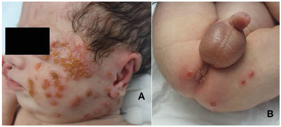
On the first day of life, a male infant born at 36 weeks’ gestation (weight 2,520 g, height 46 cm), with an antenatal diagnosis of Down’s syndrome and atrioventricular duct defect, showed hepatomegaly, splenomegaly, bradycardia and sleep apnoea. On day 1, his white blood cell count was high (50,100/mm3; normal range 10,000–26,000/mm3, with 14% polynuclear neutrophils, 20% lymphocytes, 1% monocytes and 65% blast cells), with 377,000/mm3 platelets. Immunophenotyping of peripheral blood showed a myeloblastic leukaemia profile (CD33+, CD117+, CD71+, CD7+, CD4+ and partially CD41a), and bone marrow aspiration findings revealed 54% blast cells with no megakaryocytes. At age 1 week, the infant began to develop erythematous vesicles and pustular lesions on his face (Fig. 1A), with some on his trunk and bottom (Fig. 1B). These lesions rapidly became crusty and oozing. Laboratory results found no antinuclear, anti-basal membrane or anti-intercellular substance antibodies. Bacterial, viral and fungal cultures of the pustules were negative. Histopathology of 2 cutaneous biopsies, including immunostainings for CD34, CD68P, CD45, CD1a, C-KIT and D2-40, showed polymorphous dermal inflammation and no blast cells. Topical and systemic antibiotics (mupirocin and cefazolin, respectively) for 10 days did not induce improvement.
What is your diagnosis? See next page for answer.
Fig. 1. Pustules and crusts on (A) the face and (B) gluteal area.
Acta Derm Venereol 2021; 101: adv00394.
Diagnosis: Vesiculopustules linked to transient leukaemoid reaction
Children with Down’s syndrome (trisomy 21) have increased risk of myeloproliferative disorders, and almost 10% of neonates show a leukaemoid reaction (also called transient leukaemia). In the present case, blood cell counts progressively returned to normal during the first 6 weeks of life, without treatment for leukaemia. Cutaneous lesions associated with leukaemoid reactions, as in our case, are very different from specific cutaneous manifestations called leukaemia cutis, that present as congenital skin-infiltrated macules or nodules (“blueberry muffin” syndrome) and where microscopy examination shows epidermal infiltrates of blast cells. In newborns, pustules suggest first a skin infection (bacterial, viral or fungal); however, aseptic pustules might also occur. A first case of a vesiculopustular eruption in an infant with Down’s syndrome and transitory neonatal leukaemia was published in 1996 (1). On histological examination, epidermal immature myeloid cells, inflammation and spongiosis were seen, but skin lesions spontaneously resolved as soon as peripheral blood smear findings normalized. Since then, other cases have been described, with various histological findings and mechanisms suggested (2).
In the current case, skin samples were poorly contributive. Distinctive haematological features resolved at age 6 weeks and skin lesions at age 2 months, after 10 days of daily topical steroids (diflucortolone valerate 0.1%). At age 10 months, the child showed no clinical or biological recurrence. However, laboratory tests must be repeated because myelodysplasia might further develop (3).
To conclude, newborns with a leukaemoid reaction in the peripheral blood might present aseptic vesicles and pustules, especially on the face. Bacterial, viral and fungal cultures, as well as skin biopsy, must be performed to rule out diagnoses of skin infections and leukaemia cutis. Vesiculopustular eruption progressively resolves with regression of leukaemoid reaction. Topical steroids might be applied to enhance removal of skin lesions.
The authors have no conflicts of interest to declare.


 Click to show fullsize