


1Department of Dermatology, Shinshu University School of Medicine, 3-1-1 Asahi, Matsumoto, Nagano 390-8621, 2Institute for Melanin Chemistry, Fujita Health University, Toyoake and 3Department of Physical Therapy, Shinshu University School of Health Sciences, Matsumoto, Japan. *E-mail: akn@shinshu-u.ac.jp
#These authors contributed equally to the present study.
Accepted Jan 26, 2021; Epub ahead of print Feb 1, 2021
Acta Derm Venereol 2021; 101: adv00387.
doi: 10.2340/00015555-3757
Nail apparatus melanoma (NAM) is a rare variant of cutaneous melanoma occurring in the nail unit of the fingers and toes (1). It is challenging to distinguish early-stage NAM from benign nail streak (BNS) due to melanocytic naevus, drug-induced nail pigmentation, ethnic melanonychia, or other causes, since both conditions clinically present linear brown-to-black nail pigmentation. Although biopsy of the nail matrix is essential for a definitive diagnosis, it may cause permanent deformity of the nail plate. Additional non-invasive diagnostic methods for early NAM lesions prior to biopsy are therefore required in the clinical setting.
This study aimed to determine the values of pyrrole-2,3,5-tricarboxylic acid (PTCA; a degradation product of eumelanin), thiazole-2,4,5-tricarboxylic acid (TTCA; a degradation product of benzothiazole-type pheomelanin), 4-amino-3-hydroxyphenylalanine (4-AHP; a degradation product of benzothiazine-type pheomelanin) and melanin in the nail plate, and to examine the utility of those substances as non-invasive diagnostic markers for NAM.
This study was approved by the ethics board of Shinshu University School of Medicine, Nagano, Japan. All subjects provided written consent for participation.
Data for adult patients with nail streaks who were referred to the Department of Dermatology at Shinshu University Hospital between November 2014 and December 2018 were analysed retrospectively. Patients exhibiting nail streaks on multiple fingers/toes were excluded under the assumption of a different pathogenesis. All samples were cut from the nail tip in a non-invasive and painless way for obtaining square-shaped specimens several millimetres in length and width. The values of PTCA, TTCA, and 4-AHP in the nail fragments were measured using the alkaline hydrogen peroxide oxidation method (PTCA and TTCA) or reductive hydroiodic acid hydrolysis (4-AHP) with the HPLC microanalytical method (2, 3). The total melanin in each nail sample was calculated using the previously described conversion factor (PTCA×38 + TTCA×34 + 4-AHP×9) (4). NAM was definitively diagnosed by histopathological findings (1). The diagnosis of BNS was based on clinical and dermoscopic findings and confirmed by clinical course observations (5).
Mann–Whitney U tests were employed for comparing degradation product and total melanin distribution between NAM and BNS cases. Student t-tests were adopted for comparisons of mean degradation product and total melanin values between the groups. A p-value of < 0.05 was considered statistically significant. All statistical analyses were performed using EZR version 1.40 (EZRsetup.exe) (6).
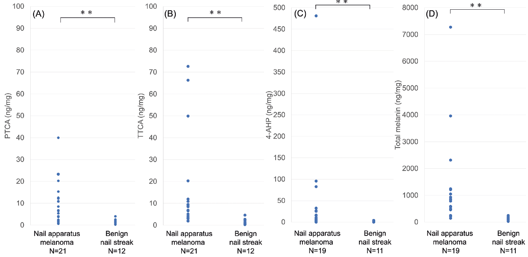
A total of 33 cases were enrolled in this study; 21 NAM cases and 12 BNS cases. The 21 NAM cases were 12 males and 9 females; median age 67 years (range 30–89 years), comprising 14 fingers and 7 toes; Breslow’s tumour thickness in situ: 12 cases; ≤ 1.0 mm: 3 cases; 1.1–2.0 mm: 4 cases; and 2.1–4.0 mm: 2 cases. The 12 BNS cases were 1 male and 11 females; median age 49 years (range 17–81 years); comprising 7 fingers and 5 toes. The distributions of PTCA, TTCA, 4-AHP, and total melanin were all significantly higher for NAM than for BNS (all p < 0.001) (Fig. 1). The mean values (ng/mg) of PTCA (9.2 vs 1.7, p = 0.019), TTCA (15.1 vs 1.7, p = 0.0079), 4-AHP (43.2 vs 0.95, p = 0.11), and total melanin (1298 vs 125, p = 0.0076) tended to be higher for in situ and invasive NAM than for BNS, with significance for PTCA, TTCA, and total melanin. Comparing in situ NAM and BNS, the mean values (ng/mg) of PTCA (11.1 vs 1.7, p = 0.019), TTCA (10.2 vs 1.7, p = 0.044), 4-AHP (24.3 vs 0.95, p = 0.045), and total melanin (981 vs 117, p = 0.024) were all significantly higher for NAM. The sensitivity and specificity for detecting in situ and invasive NAM or in situ NAM at a threshold of 5.0 ng/mg for PTCA, TTCA, 4-AHP, or any of the 3 substances or at a threshold of 405 ng/mg for total melanin are shown in Table I. An increase in any of the 3 degradation products (> 5.0 ng/mg) or total melanin (> 405 ng/mg) had excellent specificity for in situ NAM detection.
Fig. 1. Distributions of substance values in nail samples from nail apparatus melanoma and benign nail streak cases. (A) Pyrrole-2,3,5-tricarboxylic acid (PTCA). (B) Thiazole-2,4,5-tricarboxylic acid (TTCA). (C) 4-Amino-3-hydroxyphenylalanine (4-AHP). (D) Calculated total melanin. All substances were significantly higher for nail apparatus melanoma cases than for benign nail streak cases. The data for one case each of invasive nail apparatus melanoma, in situ nail apparatus melanoma, and benign nail streak could not be obtained for 4-amino-3-hydroxyphenylalanine due to an insufficient nail sample size. **p < 0.001.
Table I. Sensitivity and specificity of melanin degradation product and calculated total melanin values in nail samples for detecting nail apparatus melanoma (NAM)
This study found that PTCA, TTCA, 4-AHP, and calculated total melanin in the nail plate were significantly higher in NAM than in BNS, in adult cases. This report supports the utility of melanin degradation products as possible non-invasive indicators for NAM detection with good sensitivity and specificity. Moreover, as those values were already elevated for in situ NAM, this study method may also be applicable for detecting NAM at an early stage.
Mammals and birds produce 2 chemically distinct types of melanin (eumelanin and pheomelanin), based on which hair, skin, and eye colours are determined (7). As each melanin type is chemically synthesized in separate pathways (8), their degradation product values in cells or tissues are measured individually for quantity and ratio assessments. PTCA, TTCA, and 4-AHP are all major melanin degradation products and their quantification methods have been well established (2, 3). Although the mechanism for melanin and melanin-associated product increases is not fully understood, several studies have demonstrated elevated substance levels in melanoma samples (9–15). Among those, serum 5-S-cysteinyldopa, a precursor of pheomelanin, most sensitively increased with melanoma progression (9–12), followed by 4-AHP, a degradation product of pheomelanin (14). In contrast, the serum and urinary levels of PTCA, a degradation product of eumelanin, were at most only double in melanoma patients compared with control subjects (15).
In this study, the PTCA/TTCA and PTCA/4-AHP ratios in the nail plate varied among NAM cases and no remarkable predominance was detected (data not shown). The results also confirmed that the values of all substances were < 2 ng/mg in nail samples from 4 amelanotic NAM cases (data not shown). Together with those findings, the observed high values of melanin degradation products in NAM nail samples may reflect the quantitative changes in melanin production, but not the qualitative changes involving the eumelanin/pheomelanin ratio. Indeed, it was found that the levels of melanin degradation products were all > 5 ng/mg in 4 paediatric BNS cases, with mean scores (ng/mg) of 27.3 for PTCA, 78.9 for TTCA and 16.6 for 4-AHP.
The main limitations of this study were the small sample size and retrospective design. Larger prospective investigations, including children, adolescents, and adults, are needed to validate the utility of melanin degradation products for NAM detection.
In conclusion, melanin degradation products in the nail plate are possible biomarkers for NAM detection. The diagnostic method used in this study is non-invasive and appears to be applicable even for in situ NAM lesions.
This study was partly supported by an SGH Cancer Research Grant.
The authors have no conflicts of interest to declare.


 Click to show fullsize
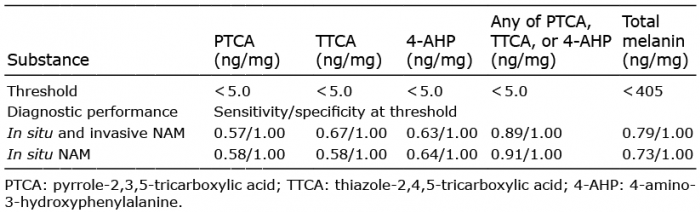 Click to show fullsize