


Woo Jin Lee1, Kwang Hee Won1, Chong Hyun Won1, Sung Eun Chang1, Jee Ho Choi1, Kee Chan Moon1, Chan-Sik Park2, Jooryung Huh2, Cheolwon Suh3 and Mi Woo Lee1
Departments of 1Dermatology, 2Pathology, and 3Oncology, Asan Medical Center, University of Ulsan College of Medicine, Seoul, Korea
Diffuse large B-cell lymphoma (DLBCL) can be separated into 2 groups: nodal and extranodal disease. The aim of this study was to analyse the clinical features of skin lesions and survival outcomes of cutaneous DLBCL according to the primary tumour site. A total of 44 patients with cutaneous DLBCL were classified as primary cutaneous DLBCL, leg type or cutaneous DLBCL secondary to primary disease. Although skin lesion characteristics did not differ significantly between groups, extensive cutaneous lesions were more often observed in secondary cutaneous DLBCL compared with DLBCL, leg type. Secondary cutaneous DLBCL was more commonly associated with an advanced stage and higher International Prognostic Index score than DLBCL, leg type. DLBCL, leg type demonstrated a better survival outcome than secondary cutaneous DLBCL. The multiplicity of skin lesions and time-point of cutaneous involvement were associated with prognosis in secondary cutaneous DLBCL. Survival outcomes and prognostic factors differ depending on the primary tumour site of cutaneous DLBCL. Key words: diffuse large B-cell lymphoma; skin; survival outcome.
Accepted May 11, 2015; Epub ahead of print May 27, 2015
Acta Derm Venereol 2015; XX: XX–XX.
Mi Woo Lee, Department of Dermatology, Asan Medical Center, University of Ulsan College of Medicine, 388-1 Pungnap-dong, Songpa-gu, Seoul 138-736, Korea. E-mail: jijulee@hanmail.net
Diffuse large B-cell lymphoma (DLBCL) can be separated into 2 groups, nodal and extranodal disease, depending on the primary site (1–3). Genetic and phenotypic differences between nodal and extranodal DLBCL have been suggested, including single-gene alterations in genes such as c-MYC, Bcl-6, REL and FAS (4–6). Approximately one-third of patients with DLBCL present with extranodal involvement (7). The most common primary extranodal sites of DLBCL are the gastrointestinal tract, head and neck, and skin/soft tissue. In a large cohort of 25,992 patients with DLBCL, primary skin/soft tissue involvement accounted for 3.3% of all cases (7).
The classification of primary cutaneous DLBCL was introduced to designate a specific subtype of lymphoma with characteristic clinicopathological, immunohistochemical, and evolutive features. Primary cutaneous DLBCL is primarily defined on the basis of morphological features of confluent sheets of large cells (i.e. centroblasts and immunoblasts) with round nuclei, irrespective of Bcl-2 expression and skin lesion location (8–10). In the new World Health Organization/European Organisation for Research and Treatment of Cancer (WHO/EORTC) classification of DLBCL, leg type is defined as a cutaneous B-cell lymphoma with a predominance of large round cells that are positive for Bcl-2 (10). However, the role of Bcl-2 expression in classifying these lymphomas remains unclear, and no difference in survival have been observed between Bcl-2-positive and -negative primary cutaneous DLBCL (8, 9). Some authors classified all cases with the characteristic round cell morphological features within the group of DLBCL, leg type, irrespective of Bcl-2 expression (8, 9, 11).
Depending on the primary site of involvement, DLBCL with cutaneous involvement can be divided into 2 distinct subsets: (i) primary cutaneous DLBCL, which initially presents in the skin, and (ii) DLBCL accompanied by secondary spread to the skin. Whereas primary cutaneous DLBCL, leg type has been widely evaluated, there are limited data available to describe the clinical features or survival analysis of patients with secondary cutaneous DLBCL. Differences in clinical features and prognosis depending on primary sites of cutaneous DLBCL have not been analysed. In the current study cutaneous DLBCL involvement was classified according to the primary tumour site, and the clinical characteristics and survival outcomes were analysed and compared between groups.
MATERIALS AND METHODS
Study population
Following approval from the Institutional Review Board of our institution, we searched the Asan Medical Center database for cases of cutaneous DLBCL that had been confirmed by skin biopsy between January 1998 and October 2013. DLBCL was defined based on a predominance (≥ 80%) of large cells with morphological features of centroblasts and immunoblasts, irrespective of Bcl-2 expression and skin lesion location. Cases showing a large proportion of large centrocytic cells (cleaved cells) were excluded. Primary cutaneous DLBCL, leg type was defined irrespective of Bcl-2 expression and skin lesion location according to previous reports (8, 9, 11). Bcl-2 and MUM-1 expression were considered positive when over 50% of tumour cells expressed these proteins.
Cutaneous DLBCL patients were classified as either primary cutaneous DLBCL, leg type (group A) or secondary cutaneous DLBCL with primary nodal or extranodal disease (group B). Primary cutaneous DLBCL, leg type was defined by skin-only disease at initial staging without evidence of extracutaneous disease. Staging was performed using standard methods at the time of initial diagnosis, including a physical examination, blood cell counts, chest radiography, computed tomographic scanning, and bone marrow biopsy. Lymph node biopsy was performed when adenopathies were clinically detectable.
Variables of interest
The following clinical data were collected from the patient medical records: age at diagnosis; sex; location, number, and morphology of skin lesions; complete blood analysis; serum lactic dehydrogenase (LDH) level; Ann Arbor stage; bone marrow biopsy; computed tomographic imaging; presence of B symptoms; International Prognostic Index (IPI) score; type of treatment; follow-up results; and survival. IPI scores were calculated using each of the following risk factors: age > 60 years, stage III or IV disease, elevated serum LDH, ECOG performance status of 2, 3, or 4, and more than one extranodal site (12). The degree of skin involvement was evaluated using the multiplicity and extent of cutaneous lesions. The number of skin lesions was grouped as single or multiple (≥ 2 lesions). The degree of the extent of the cutaneous lesions was classified as “localized” when one or multiple skin lesions were restricted to one anatomical site and “extensive” when several non-contiguous anatomical sites according to ISCL-EORTC classification were involved (13). The time-point of cutaneous dissemination in secondary cutaneous DLBCL was defined as “early” when skin lesions were noted within 6 months after the initial diagnosis and “late’’ when they developed ≥ 6 months after the initial diagnosis.
Overall survival (OS) was calculated from the date of initial diagnosis to the date of death from any cause or the last follow-up examination. Progression-free survival (PFS) was calculated from the date of the initial diagnosis to the first day of disease progression, relapse, or last follow-up. Prognostic factors associated with survival were evaluated using clinical data at the time of initial staging.
Statistical analysis
Survival analysis was performed according to the Kaplan–Meier method, and significance was tested using the log-rank test. Prognostic factors at the time of diagnosis independently associated with OS were identified by multivariate analysis using Cox proportional hazards regression modelling. Comparisons between subgroups of patients according to primary tumour site were performed using a χ2 test or Fisher’s exact test for categorical variables and a t-test or Mann–Whitney test for continuous variables. All analyses were performed using a statistical software package (SPSS, version 18.0; SPSS Inc., Chicago, IL). A p-value < 0.05 was considered statistically significant.
RESULTS
A total of 44 cases of DLBCL with skin involvement were identified among 2,067 cases of DLBCL through a retrospective review of the medical database of Asan Medical Center. The clinical characteristics of these patients are summarized in Table SI1. The initial sites of presentation were the skin (primary cutaneous DLBCL, leg type, group A; n = 14) or the lymph node or other extranodal sites except for the skin (secondary cutaneous DLBCL, group B; n = 30). The lymph node was the most common primary tumour site in secondary cutaneous DLBCL (Table SII1). Of 30 patients with secondary cutaneous DLBCL, 17 developed dissemination on sites other than the skin when cutaneous involvement was found. Extracutaneous involvement in DLBCL, leg type was noted in 8 (57%) out of 14 patients (Table SII1) and the median time until extracutaneous involvement after initial diagnosis was 18.4 months (range 7–48 months).
Clinical findings according to primary tumour site
When all DLBCL patients were combined into a single cohort and analysed, the secondary cutaneous DLBCL group showed a higher proportion of patients with disease dissemination (Table SI1). An advanced stage (p = 0.000), high (> 2) IPI score (p = 0.000), B symptoms (p = 0.049), and elevated LDH (p = 0.025) were significantly more common for secondary cutaneous DLBCL than for DLBCL, leg type.
Clinical features and immunophenotypic profile of cutaneous lesions (Table SI1)
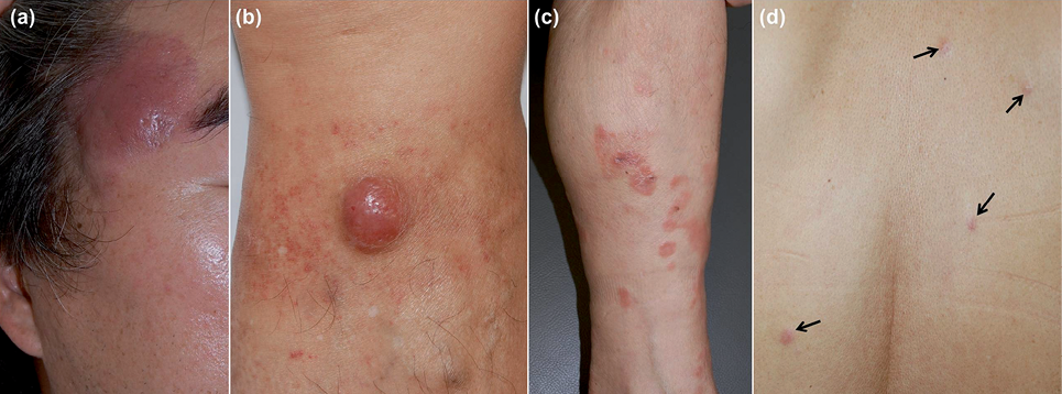
The leg was the most commonly involved site in both groups. The initial skin lesion in primary cutaneous DLBCL, leg type was located at the following sites: legs (n = 6), scalp (n = 2), back (n = 2), abdomen (n = 2), face (n = 1), and arm (n = 1). The clinical presentation consisted of cutaneous nodules or tumours (21/44, 47%), subcutaneous nodules (15/44, 34%), papular lesions (7/44, 16%), and indurated plaques (3/44, 6.8%) (Fig. 1). Although no statistically significant difference with respect to the multiplicity of skin lesions was observed (p = 0.276), extensive skin lesions were more common in group B than in group A (p = 0.010). In the immunohistochemical study, no significant differences were found for Bcl-2 and Bcl-6 expression between the 2 groups.
Variables in cutaneous lesions
Primary cutaneous DLBCL, leg type. The degree of skin involvement, including multiplicity (p = 0.881) and extent (p = 0.498), had no significance effect on the development of extracutaneous involvement. A leg location in group A did not predict either clinical features of skin lesions, such as multiplicity (p = 0.254) and extent (p = 0.475), or extracutaneous involvement (p = 0.315).
Secondary cutaneous DLBCL. Cutaneous dissemination was noted between 0 and 46 months (median 11 months) after the initial diagnosis of the primary tumour. Multiple skin lesions were more common in patients who developed cutaneous dissemination within 6 months after the initial diagnosis of the primary tumour than in patients with cutaneous dissemination 6 months or more after the initial diagnosis in group B (p = 0.045). Location on the leg did not predict clinical features of skin lesions, such as the multiplicity (p = 0.815), extent (p = 0.149), and time-point (p = 0.293) of skin lesion development. Clinical features (multiplicity, extent, morphology, time-point, and location) of skin lesions were not significantly different depending on primary tumour site (nodal vs. extranodal) in group B.
Survival outcomes in cutaneous diffuse large B-cell lymphoma
Overall, 30 patients (68%) received chemotherapy only, and 8 (18%) and 3 (6.8%) patients were treated with chemotherapy with radiotherapy or chemotherapy with autologous bone marrow stem cell transplant, respectively. The most common chemotherapy regimen was R-CHOP (rituximab, cyclophosphamide, doxorubicin, vincristine, and prednisone; 24/44, 55%), followed by CHOP (14/44, 32%) and ESHAP (etoposide, methylprednisolone, cytarabine, and cisplatin; 8/44, 18%). Follow-up periods ranged from 1 to 144 months (median follow-up, 41 months). Of 44 patients in the study cohort, 19 (43%; 7 patients in group A and 12 patients in group B) died from disease progression, after a period of between 2 and 68 months (group A: 7–68 months, median 39 months; group B: 2–60 months, median 17 months). Twenty patients (45%) are alive with or without disease, and the remaining 5 patients (11%; 2 patients in group A and 3 patients in group B) were lost to follow-up. When all patients were combined into a single cohort, the 5-year OS rate was 43% and the median OS period was 42.0 months (95% confidence interval (95% CI): 19.83–63.17 months). Patients with DLBCL, leg type demonstrated significantly more favourable survival outcomes than patients with secondary cutaneous DLBCL (Fig. S1a1: OS; Fig S1b1: PFS). The values of the median OS, 5-year OS rate, and median PFS in group A were higher than those in group B (Table I).
Table I. Survival data of 47 patients with cutaneous diffuse large B-cell lymphoma (DLBCL)
|
Median OS (95% CI), months |
5-year OS rate, % |
Median PFS (95% CI), months |
|
|
Total |
42.0 (19.83–63.17) |
43 |
21.0 (8.12–40.82) |
|
Group Aa |
67.0 (39.29–91.23) |
65 |
40.0 (13.17–70.83) |
|
Group Bb |
30.0 (15.23–44.77) |
31 |
16.0 (7.26–26.23) |
|
p-value |
0.047* |
0.025* |
|
|
Variables in group Aa |
Median OS (95% CI), months |
||
|
Extent of skin lesions |
Extensive skin lesions |
11.0 (**) |
|
|
Localized skin lesions |
67.0 (43.12–85.74) |
||
|
p-value |
0.023* |
||
|
Extracutaneous involvement |
Positive |
37.0 (21.42–59.54) |
|
|
Negative |
67.0 (**) |
||
|
p-value |
0.042* |
||
|
Location of skin lesions |
Leg site |
39.0 (**) |
|
|
Non-leg sites |
69.0 (**) |
||
|
p-value |
0.027* |
||
|
Variables in group Bb |
Median OS (95% CI), months |
Median PFS (95% CI), months |
|
|
Multiplicity of skin lesions |
Multiple |
19.0 (14.71–23.69) |
10.0 (6.93–13.71) |
|
Single |
33.0 (23.14–42.12) |
21.0 (18.24–25.67) |
|
|
p-value |
0.037* |
0.071 |
|
aGroup A: DLBCL, leg type. bGroup B: secondary cutaneous DLBCL.
*Statistically significant.
**No data were found.
OS: overall survival; PFS: progression-free survival; CI: confidence interval.
Primary cutaneous diffuse large B-cell lymphoma, leg type (Group A)
Patients with extensive skin lesions demonstrated a poorer median OS than patients with localized skin lesions (p = 0.023, Table I). In contrast, the multiplicity of skin lesions did not predict the OS of patients with DLBCL, leg type (p = 0.287). Patients with extracutaneous lesions secondary to DLBCL, leg type demonstrated a poorer median OS (p = 0.042, Table I). Cutaneous lesions located on the legs were also associated with poor clinical outcomes (p = 0.027, Table I). In multivariate analysis, no independent prognostic factors were found.
Secondary cutaneous diffuse large B-cell lymphoma (Group B)
The survival outcomes of patients with single skin lesions were significantly different from those of patients with multiple skin lesions (Fig. S1c1: OS; Fig. S1d1: PFS; Table I). In contrast to the results found for the multiplicity of skin lesions, extensive lesions did not affect OS in patients with secondary cutaneous DLBCL (p = 0.274). The time-point of cutaneous involvement affected the OS outcomes. Patients with cutaneous involvement within 6 months after the initial diagnosis of primary disease demonstrated worse survival outcomes than patients who developed cutaneous dissemination 6 months or more after the initial diagnosis (Fig. S1e1: OS; Fig. S1f1: PFS; Table I). Multivariate analysis of OS using all candidate variables identified an early time-point of cutaneous dissemination (hazard ratio 1.09; 95% CI 1.01–5.95; p = 0.047) as an independent factor associated with a poorer prognosis.
DISCUSSION
Primary cutaneous lymphomas may behave very differently from their nodal counterparts. For example, there may be differences in genetic aberrations and the clinical course, in that localized cutaneous large B-cell lymphoma follows a less aggressive clinical course compared with large B-cell lymphoma arising from nodal sites (14–16). Primary cutaneous DLBCL, leg type is characterized by a predilection for the leg, a high proportion of Bcl-2 expression, frequent relapses, and extracutaneous dissemination (8). The present study included primary DLBCL, leg-type cases, based on morphological characteristics, irrespective of Bcl-2 positivity, because there have been controversies over the role of Bcl-2 expression and the location of skin lesions in the classification of DLBCL, leg type (8–11). Between approximately 70% and 80% of DLBCL, leg type patients have lesions on the legs (8, 17, 18), but there are limited data available to describe the clinical characteristics of primary cutaneous DLBCL in Asians. The present study revealed that 6 of 14 patients (43%) with primary cutaneous DLBCL, leg type had skin lesions on the legs. Primary DLBCL, leg type is a rare cutaneous lymphoma entity in Asians. The reason for the different anatomical distribution in Asians in the present study is unclear, but may reflect racial differences.
The cutaneous lesions in the present study demonstrated clinical aspects that are similar to those of cases reported previously in the literature (8, 17–22). The clinical presentations varied from cutaneous nodules to papules or infiltrated plaques, but the most common manifestation was cutaneous nodules on the legs. There were no significant differences between our patient groups in terms of the anatomical location, multiplicity, and morphology of the skin lesions, except for the extent of the lesions. Extensive cutaneous involvement in several non-contiguous anatomical sites was more common in secondary cutaneous DLBCL, but multiplicity of skin lesions was not significantly different between the groups. Clinical features (multiplicity, extent, location on the leg) of skin lesions in DLBCL, leg type did not predict the development of extracutaneous involvement. Although skin lesions on the leg were associated with a worse clinical outcome, the skin lesion location in DLBCL, leg type had no significant effect on the characteristics (multiplicity, extent, and morphology) of the skin lesions in the present study. However, the significance of these results could be limited by the small number of DLBCL, leg-type cases in the present study.
Patients with DLBCL vary in clinical presentation and prognosis, show variable response rates to standard chemoimmunotherapy, and have 5-year OS rates ranging from 30% to 80% (7, 23, 24). Therapeutic responses and prognosis seem to differ depending on the DLBCL subgroup (24–27). Primary extranodal sites of involvement seem to be associated with distinct outcomes in patients with DLBCL (7, 28). Although OS in patients with primary extranodal involvement was revealed to be significantly higher than that of the primary nodal group (28), specific sites of involvement seemed to be associated with either a better or a worse prognosis (7, 14–16).
The 5-year survival rate in secondary cutaneous DLBCL in the present study (31%) was lower than that of previous reports (7, 23, 24, 29). This difference in survival may be because “secondary cutaneous DLBCL” only included patients with systemic DLBCL with cutaneous dissemination, leading to differences in the baseline characteristics of patients. Cutaneous involvement secondary to systemic DLBCL would suggest disease progression and poorer outcomes than conventional DLBCL. The 5-year survival in DLBCL, leg type was 65%, which is in accordance with the data in the literature reporting 5-year survival rates of 37% to 67% (8, 9, 11, 17, 30). The survival of cutaneous DLBCL patients differed depending on the primary tumour site. DLBCL, leg type was more likely to be less aggressive and demonstrate better prognosis than secondary cutaneous DLBCL with systemic disease. This result is in agreement with that of a previous study that reported that primary extranodal DLBCL has a better prognosis than nodal DLBCL (28). Although it was not statistically significant, primary cutaneous DLBCL was associated with better survival than nodal DLBCL (7).
Various poor prognostic factors for DLBCL have been reported in previous studies. Unfavourable variables predicting OS in DLBCL include age older than 60 years, B symptoms, poor performance status, advanced stage, extranodal involvement, bone marrow involvement, high serum LDH levels, and high β2m and Bcl-2 protein expression (24, 28, 29, 31). In primary cutaneous DLBCL, a leg location and multiple skin lesions were indicative of worse prognosis (8, 9, 18). The 3-year disease-specific survival rates were 39% and 77% in patients with multiple skin lesions and single lesions, respectively (8). To our knowledge, there has been no prognostic analysis in patients with secondary cutaneous DLBCL with systemic disease.
The degree of skin involvement significantly affected prognosis in both primary and secondary cutaneous DLBCL. However, there was a small difference in the effect of the degree of skin involvement on prognosis depending on the primary site of cutaneous DLBCL. Extensive cutaneous lesion in several anatomical sites was associated with poor prognosis only in primary cutaneous DLBCL, leg type, whereas the number of skin lesions (multiplicity) had a significant effect on prognosis only in secondary cutaneous DLBCL. Location on the leg was indicative of worse prognosis in DLBCL, leg type but had no prognostic significance in patients with secondary cutaneous DLBCL. Half of the patients with DLBCL, leg type in the present study developed extracutaneous involvement, and this was associated with poor survival in comparison with patients who did not develop extracutaneous involvement. In the present study, 4 of 7 patients with DLBCL, leg type who died presented with skin lesions on the legs and developed extracutaneous dissemination.
Prognosis of secondary cutaneous DLBCL was related to the interval from the date of initial diagnosis to the development of skin lesions. Secondary cutaneous involvement soon after the initial diagnosis of primary disease predicted more aggressive clinical outcomes and was more likely to present with multiple skin lesions than secondary cutaneous involvement at a later time after initial diagnosis. Prognostic factors in conventional DLBCL, such as serum LDH, stage, age, sex, primary site of secondary cutaneous DLBCL (nodal vs. extranodal sites), and IPI score, had no prognostic significance in secondary cutaneous DLBCL. Secondary cutaneous involvement in systemic DLBCL suggests that the patient is at high risk of disease progression and poor prognosis, which is not affected by the prognostic factors of conventional DLBCL.
There were some limitations to our study due to its retrospective design. Age (> 70 years) at time of diagnosis has been known to be significantly associated with worse prognosis in primary cutaneous DLBCL, leg type (8, 9, 18), but our present analyses could not validate this association because only 3 patients older than 70 years were included. Although a previous study demonstrated that Bcl-2 expression and absence of Bcl-6 expression predicted poor survival in systemic DLBCL (6, 24, 32), no significant difference in survival was noted according to antigen expression in both groups from our current study. In our present analysis, positivity for Bcl-2 in DLBCL, leg type had no effect on survival, in accordance with the results of previous reports (8, 9, 17). However, there is an inherent limitation resulting from the small sample size of immunostainings.
In conclusion, the prognostic factors that influence survival differed depending on the primary tumour site of cutaneous DLBCL. Secondary cutaneous DLBCL demonstrated extensive skin lesions that involved multiple anatomical sites and showed poorer OS than DLBCL, leg type. Extensive cutaneous lesions and cutaneous involvement soon after initial diagnosis predicted a poorer OS in patients with secondary cutaneous DLBCL.
The authors declare no conflicts of interest.

1http://www.medicaljournals.se/acta/content/?doi=10.2340/00015555-2139
REFERENCES


