


Alexander Kreuter, Peter Altmeyer and Thilo Gambichler
Department of Dermatology and Allergology, Ruhr-University Bochum, Gudrunstrasse 56, DE-44791 Bochum, Germany. E-mail: a.kreuter@derma.de
Accepted November 11, 2005.
Sir,
Primary cutaneous CD30+ anaplastic large cell lymphoma (ALCL) is defined by the following criteria: (i) predominance or large clusters of CD30+ blast cells in the initial skin biopsy specimen; (ii) clinically no evidence of lymphomatoid papulosis; (iii) no prior or concurrent lymphomatoid papulosis (LyP), mycosis fungoides (MF), or other type of cutaneous lymphoma or extracutaneous localization at presentation (1, 2). Lesions of primary cutaneous CD30+ ALCL tend to be large nodules, tumours, or subcutaneous masses that often ulcerate. Primary cutaneous CD30+ lymphoproliferative disorders, including CD30+ ALCL, LyP and borderline cases, represent a clinical and histological continuum. In contrast to primary systemic and secondary CD30+ ALCL (e.g. large-cell transformation of MF), the primary cutaneous variant is characterized by an indolent clinical course with 5- and 10-year survival rates exceeding 95% and disseminated or extracutaneous disease in less than 10% (1).
There is no uniform therapeutic approach for CD30+ ALCL, but most patients receive combinations of surgery, local radiation and/or chemotherapy (2, 3). Bexarotene, a retinoid X-receptor (RXR)-selective retinoid, has recently been approved by the Food and Drug Administration (FDA) for cutaneous T-cell lymphoma including refractory MF and Sézary syndrome (4). There are only anecdotal reports on the treatment of CD30+ ALCL with bexarotene (5, 6).
We describe here a patient with a predominantly intertriginous manifestation of primary cutaneous ALCL, who rapidly developed extracutaneous tumour mass following bexarotene and interferon-α (IFN-α).
CASE REPORT
A 41-year-old Caucasian woman was referred to our hospital with a 10-month history of progressive cutaneous tumours located mainly in the intertriginous areas such as the axillae, submammary region and inguinal folds (Fig. 1). Apart from slight pruritus she felt otherwise healthy, and had no concomitant diseases except obesity. Several skin biopsies showed mainly dermal and in part subcutaneous infiltration of atypical large lymphoid cells with pleomorphic nuclei and prominent nucleoli. Immunophenotyping revealed that these cells were strongly positive for CD30, CD4 and CD3, and negative for CD20 and CD56. A complete work-out, including clinical and bone marrow examinations, sonography and computerized tomography (CT), showed no evidence for systemic disease. A diagnosis of primary cutaneous CD30+ ACLC was made. Surgical treatment was not considered because of the number of skin lesions. Hence, local radiotherapy with a total dose of 40 Gy given as 20 daily 2-Gy fractions was performed, resulting in a marked decrease of tumour load. However, she experienced new lesions shortly after finishing radiotherapy, and therefore monthly cycles of extracorporeal photophoresis (ECP) in combination with 3×4.5 MIU/week IFN-α-2b were initiated. After a stable outcome for 9 months, new lesions appeared, so bexarotene was added at a dosage of 300 mg/m2 per day. Two months later increasing anaemia and lymphopaenia forced us to discontinue ECP and reduce IFN-α (3×3 MIU/week). Six months after beginning bexarotene, she presented with distinctive swelling of her right leg. A CT scan of the pelvis revealed a tumour mass of 9×7.5×9 cm infiltrating both arteria and vena femoralis communis (Fig. 2). CT-guided fine needle biopsy confirmed infiltration of T-cell lymphoma. However, these tumour cells were no longer positive for CD30 and CD4, but only for CD3. Otherwise there was no evidence for systemic disease. Bexarotene and IFN-α treatment were discontinued. Flow cytometric blood analysis before and after initiation of bexarotene did not reveal pathologies for CD3, CD4, CD7, CD26, CD56 and CD30 expression of lymphocytes. Multi-agent chemotherapy with ifosfamide, etoposide, vincristine, and dexamethasone followed by cyclophosphamide, epirubicine, and dexamethasone was initiated for two cycles. However, disease progressed and chemotherapy was changed to cisplatin, cytarabine and dexamethasone. The patient died of severe sepsis and tumour cachexia 5 months after onset of extracutaneous spread.
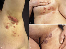
Fig. 1. Multiple nodules and plaques located in the (a) left axilla, (b) right submammary region and (c) left inguinal fold.
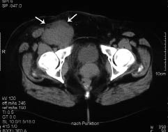
Fig. 2. Computed tomography scan of the pelvis demonstrating a bulky tumour mass (arrows), infiltrating both arteria and vena femoralis communis.
DISCUSSION
We describe here a patient with primary cutaneous CD30+ ALCL that primarily affected intertriginous areas. Apart from granulomatous slack skin, which is a distinct T-helper (Th) cell lymphoma excluded on the basis of the clinicopathological findings in our patient cutaneous lymphomas are usually not observed predominantly in these regions (2, 3). Our patient was initially expected to have a good prognosis. However, she experienced a dramatic progression of her disease following 6-months’ therapy with bexarotene and IFN-α, which has been rarely used in CD30+ ACLC (5, 6).
CD30 is preferentially expressed on CD4+ T-cell clones that produce Th2-type cytokines, such as interleukins 4 and 5. IFN-α as well as bexarotene antagonize Th2-type cytokine production by malignant cells and thus enhance the effector phase of anti-tumour immunity. Moreover bexarotene has the ability to induce apoptosis within the malignant population of cells (3). In a recent study, however, purified Sézary cells from approximately one-third of patients demonstrated significant resistance to apoptosis. One might speculate that the beneficial effects are restricted to cutaneous T-cell infiltration, whereas circulating tumour cells are relatively unaffected (7). Furthermore bexarotene has been shown to down-regulate E-selectin on endothelial cells, thus causing the entrapment of cutaneous homing T lymphocytes in the circulation (8).
So far, only one patient with primary cutaneous CD30+ ACLC has been reported to respond to bexarotene monotherapy (4). Recently, 6 patients with advanced recalcitrant CTCL, including 2 with CD30+ large-cell transformation, have been described experiencing extracutaneous disease under bexarotene therapy despite significant cutaneous improvement (6). We recently reported a heavily pre-treated Sézary patient who had a rapid onset of extracutaneous lymphoma following monotherapy with oral bexarotene (9). Our patient with CD30+ ACLC developed a bulky tumour mass in the pelvis following bexarotene/IFN-α combination therapy. A transformation of immunophenotypes of the pelvic tumour mass indicated a poor prognosis. The temporal relation prompts us to assume bexarotene alone, or possibly the combination with IFN-α, to be causally involved in extracutaneous lymphoma transformation.
REFERENCES
1. Bekkenk MW, Geelen FAM, van Voorst Vader PC, Heule F, Geerts ML, van Vloten WA, et al. Primary and secondary cutaneous CD30(+) lymphoproliferative disorders: a report from the Dutch Cutaneous Lymphoma Group on the long-term follow-up data of 219 patients and guidelines for diagnosis and treatment. Blood 2000; 15: 3653–3661.
2. Paulli M, Berti E, Rosso R, Boveri E, Kindl S, Klersy C, et al. CD30/Ki-1-positive lymphoproliferative disorders of the skin–clinicopathologic correlation and statistical analysis of 86 cases: a multicentric study from the European Organization for Research and Treatment of Cancer Cutaneous Lymphoma Project Group. J Clin Oncol 1995; 13: 1343–1354.
3. Kim EJ, Hess S, Richardson SK, Newton S, Showe LC, Benoit BM, et al. Immunopathogenesis and therapy of cutaneous T-cell lymphoma. J Clin Invest 2005; 115: 798–812.
4. Talpur R, Ward S, Apisarnthanarax N, Breuer-Mcham J, Duvic M. Optimizing bexarotene therapy for cutaneous T-cell lymphoma. J Am Acad Dermatol 2002; 47: 672–684.
5. Keung YK, Woodruff R, Sangueza O. Response of CD30+ large cell lymphoma of the skin to bexarotene. Leuk Lymphoma 2002; 43: 1153–1154.
6. Bouwhuis SA, Davis MD, el-Azhary RA, McEvoy MT, Gibson LE, Knudsen JM, et al. Bexarotene treatment of late-stage mycosis fungoides and Sezary syndrome: development of extracutaneous lymphoma in 6 patients. J Am Acad Dermatol 2005; 52: 991–996.
7. Brennand S, Sutton VR, Biagi J, Trapani JA, Westerman D, McCormack CJ, et al. Lack of apoptosis of Sézary cells in the circulation following oral bexarotene therapy. Br J Dermatol 2005; 152: 1199–1205.
8. el-Azhary RA, Bouwhuis SA. Oral bexarotene in a therapy-resistant Sézary syndrome patient: observations on Sézary cell compartmentalization. Int J Dermatol 2005; 44: 25–28.
9. Kreuter A, Altmeyer P. Rapid onset of CD8-positive aggressive T-cell lymphoma during bexarotene therapy in a patient with Sézary syndrome. J Am Acad Dermatol 2005; 53: 1093–1095.

