Karin Terstappen1, Olle Larkö2 and Ann-Marie Wennberg2
Departments of Dermatology, 1Kärnsjukhuset, Skövde, and 2Sahlgrenska University Hospital, Göteborg, Sweden


Karin Terstappen1, Olle Larkö2 and Ann-Marie Wennberg2
Departments of Dermatology, 1Kärnsjukhuset, Skövde, and 2Sahlgrenska University Hospital, Göteborg, Sweden
Pigmented basal cell carcinomas can be difficult to distinguish clinically from melanoma. Dermoscopy has proven to be useful in the differential diagnosis of the two tumour types. SIAscopy (Spectrophotometric intracutaneous analysis) is a fairly new technique of imaging pigmented skin lesions that has been presented previously as a useful tool in diagnosing melanoma. The aim of this study was to evaluate whether SIAscopy can be useful in diagnosing pigmented basal cell carcinomas. Twenty-one pigmented basal cell carcinomas were analysed, comparing dermoscopic and SIAscopic findings. The results, in this limited setting, show that SIAscopy has no advantages over dermoscopy when diagnosing pigmented basal cell carcinomas. On the contrary, pigmented basal cell carcinomas show, in SIAscopy, similar features to those previously reported for melanoma. Key words: dermoscopy; dermatoscopy; diagnosis; melanoma; pigmented basal cell carcinoma; SIAscopy.
(Accepted December 8, 2006.)
Acta Derm Venereol 2007; 87: 238–242.
Karin Terstappen, Department of Dermatology, Kärnsjukhuset, SE-541 85 Skövde, Sweden. E-mail: karin.terstappen@vgregion.se
Basal cell carcinomas (BCCs) are the most common type of malignant skin tumours, and they are composed of cells that originate from the basal cell layer of the epidermis. They very rarely metastasize. A proportion of BCCs contain pigment. In the largest histological series studied the incidence ranges from 6.7% to 8.5%, but a racial predilection probably exists (1, 2). Most of the BCCs’ histological patterns can have pigmented varieties; however, morphoeic and infiltrative subtypes are rarely pigmented. Histologically, melanin can be found in the tumour mass and surrounding dermis. Within the tumour mass, melanocytes are often hyperplastic and melanosomes are often confined to the melanocytes. However, they may be taken up by surrounding malignant epithelial cells (3, 4). Phagocytosis of melanosome-containing apoptotic cells by the neighbouring tumour cells appears to be followed by formation of melanosome complexes (5). Melanin is more often seen in the superficial component of the tumour (1). In the dermis, melanin is found primarily in melanophages, but small amounts may be lying free. This dermal pigment is typically found at the tumour shoulders, but is occasionally located below the tumour, especially when melanocytes are found in deep tumour nodules (1). Finally, hyperplastic melanocytes may be found in the overlying epidermis (1).
Because of their growth patterns and asymmetry of pigmentation, pigmented BCCs are included in the differential diagnosis of invasive melanoma. They may also be confused with other benign pigmented skin lesions.
Dermoscopy
Dermoscopy is a non-invasive diagnostic technique that uses optical magnification to examine skin lesions in situ. Dermoscopy allows the epidermis to become translucent, and therefore permits a detailed examination of the pigmented structures of the epidermis, dermo-epidermal junction and, to a lesser extent, the dermis. The result is the visualization of a multitude of morphological features, not visible to the naked eye, that enhance the clinical diagnosis of nearly all pigmented lesions (6–16).
Dermoscopy has been proven to be useful in diagnosing pigmented BCCs. Dermoscopy shows that BCCs are asymmetrical and often have large areas of hypomelanotic surface. A majority of all BCCs have < 50% of their area pigmented, and only 7% of lesions have > 75% of pigmented tumour area (17).
A simple dermoscopy method for diagnosing pigmented BCCs has been created by Menzies et al. (17). This method has a sensitivity of 93% for the diagnosis of pigmented BCCs and a specificity of 89% vs. an invasive melanoma set and 92% vs. a benign pigmented skin lesion set. With this method, for a pigmented BCC to be diagnosed, it must not have the negative feature of a pigmented network and must have one or more of the six positive features (Table I).
Table I. Method of diagnosis of pigmented basal cell carcinomas. Adopted from ref (17).
| Negative feature (cannot be found) |
| Pigment network |
| Positive features (at least 1 feature found) |
| Ulceration |
| Large blue-grey ovoid nests |
| Multiple blue-grey globules |
| Maple leaf-like areas |
| Spoke wheel areas |
| Arborizing (tree-like) telangiectasia |
The features that are pigmented in the above-mentioned method are large blue-grey ovoid nests, blue-grey globules, maple leaf-like areas and spoke wheel areas. Spoke wheel areas are well-circumscribed radial projections, usually brown, but sometimes blue or grey, meeting at an often darker central axis. Large blue-grey ovoid nests are confluent, or near confluent, pigmented ovoid or elongated areas, larger than globules, and not immediately connected to a pigmented tumour body. Maple leaf-like areas are brown to blue-grey discrete bulbous extensions forming a leaf-like pattern. Multiple blue-grey globules should be differentiated from multiple blue-grey dots (melanophages) (17).
SIAscopy
Spectrophotometric intracutaneous analysis (SIA) is a relatively new technique for imaging pigmented skin lesions. The SIAscope is a computerized equipment with a handheld unit that operates by probing the skin spectrally with radiation ranging from 400 to 1000 nm. The spectrophotometric input from the skin is analysed by computer algorithms, and this results in images giving information regarding total melanin content of the epidermis and the papillary dermis, collagen and haemoglobin content, as well as the presence of melanin in the papillary dermis. SIAscopy was presented by Moncrieff et al. as a tool to help diagnose melanoma (18). Significant findings in invasive melanoma were the presence of blood displacement with erythematous blush, collagen holes and dermal melanin within the lesion (Table II). In their study, when diagnosing invasive melanoma, SIAscopy had a sensitivity of 82.7% and a specificity of 80.1% (18). This can be compared with the results obtained in a study to evaluate a two-step procedure for the dermoscopic classification of pigmented skin lesions, where the sensitivity varied between 82.6% and 85.7%, depending on which diagnostic algorithm was used, and the specificity was 70.0−83.4% (16).
Table II. SIAscopic features defined.
Adopted from ref (18).
| Dermal melanin | The presence of dermal melanin within the lesion not due to hairs |
| Erythematous blush | A peripheral increase in blood within the lesion compared with the surrounding normal skin for three-quarters of the circumference of the lesion. |
| Blood displacement | A confluent, non-pixelated area demonstrating an absence of blood within the lesion. |
| Collagen holes | An area within the lesion coinciding with melanin (dermal or total) demonstrating an absence of collagen not due to hair follicles or sebaceous/sweat ducts |
As far as diagnosing pigmented skin lesions other than invasive melanoma with SIAscopy, nothing has been published.
Our aim in this study was to examine pigmented BCCs using SIAscopy to determine what the tumours look like with this technique. Does SIAscopy make it easier to diagnose pigmented BCCs or to differentiate pigmented BCCs from melanomas?
As pigmented BCCs, especially heavily pigmented BCCs, are quite uncommon in the Swedish population, we expected that a very limited number of patients would be included in the study; thus it can be considered a pilot study.
SUBJECTS AND METHODS
Following approval by the ethics review committee, patients were recruited from the outpatient clinics in the Dermatology departments at Sahlgenska University Hospital, Göteborg and Kärnsjukhuset, Skövde. Only patients with clinically suspicious pigmented BCCs, all above the age of 18 years, were included. Written informed consent was given by all included patients. In total 25 lesions were included. On histopathological examination 21 of the 25 lesions were found to be pigmented BCCs and these were analysed further. The other four lesions were; one melanoma, one Spitz nevus, one benign pigmented nevus and the last one only showed inflammation.
The data were collected by the main author (KT) in all but two cases (in which data were collected by CS, see acknowledgements).
All lesions were scanned using the SIAscope (Astron Clinica, Cambridge, UK), giving both dermoscopic images and SIAscopic images. The lesions were analysed by one of the authors (KT) using the dermoscopy method described by Menzies et al. (17) and the SIAscopic technique described by Moncrieff et al. (18) to measure melanin content (total and dermal) and the influence of the tumour on collagen and blood.
RESULTS
The dermoscopy and SIAscopy findings are presented in Table III. The SIAscopic findings are divided into dermal melanin (DM) and total melanin (TM), blood displacement, erythematous blush and collagen holes. The finding of melanin, total and dermal, has been subdivided to show how the different dermoscopic features present melanin.
Table III. Positive dermoscopic and SIAscopic features found in the pigmented basal cell carcinoma (BCC) included in the study
| Tumour number | Ulceration | Large blue-grey ovoid nests SIAscopy findingsa | Multiple blue-grey globules SIAscopy findingsa | Maple leaf-like areas SIAscopy findingsa | Spoke wheel areas SIAscopy findingsa | Arborizing telangiectasia | Blood displacement | Erythematous blush | Collagen holes | SIAscopic findings of melanin with unspecific dermoscopic patterna | Histopathology diagnosis |
| 1 | + | + | TM/DM | Nodular BCC | |||||||
| 2 | + (TM/DM) | + (TM/DM) | + (TM) | + | + | TM | BCC (unspecified)b | ||||
| 3 | + | + (TM/DM) | + | Superficial BCC | |||||||
| 4 | + (TM/DM) | + (TM/DM) | + (TM/DM) | + | + | TM/DM | Infiltrating BCC | ||||
| 5 | + (TM) | + (TM/DM) |
| TM | Infiltrating BCC | ||||||
| 6 | + (TM/DM) | + (TM) | + | + | TM | Nodular BCC | |||||
| 7 | + | + (TM/DM) | + | TM | Infiltrating BCC | ||||||
| 8 | + (TM/DM) | + | TM | Nodular BCC | |||||||
| 9 | + | + (TM) | + | + | TM | Nodular BCC | |||||
| 10 | + (TM/DM) | + (TM/DM) |
| + | + | + | + | TM/DM | Infiltrating BCC | ||
| 11 | + (TM/DM) | TM | Infiltrating BCC | ||||||||
| 12 | + | TM/DM | Nodular BCC | ||||||||
| 13 | + | + (TM/DM) | + |
|
| TM/DM | BCC (unspecified)b | ||||
| 14 | + (TM/DM) | + |
| + | TM | Nodular BCC | |||||
| 15 | TM/DM | Nodular BCC | |||||||||
| 16 | + | + (TM/DM) | + (TM/DM) | + | TM | Nodular BCC | |||||
| 17 | + | + (TM/DM) |
| + | + | + | TM | Nodular BCC | |||
| 18 | + | + (TM/DM) | + (TM/DM) | + | + | Nodular BCC | |||||
| 19 | + | + (TM/DM) | + | Nodular BCC | |||||||
| 20 | + (TM) | + | TM/DM | Nodular BCC | |||||||
| 21 | + (TM/DM) | TM | Superficial BCC | ||||||||
| Positive features (%) | 43 | 48c (48d/48e) | 38c (38d/33e) | 29c(29d/14e) | 14c (14d/9e) | 57 | 19 | 43 | 14 | 86d/33e |
SIAscopy: Spectrophotometric intracutaneous analysis
aSIAscopic findings of increased total melanin (TM) and showing dermal melanin (DM).
bTreated with curettage due to which growth pattern could not be determined.
cPercentage of lesions showing positive dermoscopic feature.
dPercentage of lesions showing increased TM.
ePercentage of lesions showing DM.
Pigmented features
Of the pigmented features, large blue-grey ovoid nest and blue-grey globules are mostly heavily pigmented, often showing an increase in TM as well as DM. Maple leaf-like areas are much lighter in pigmentation, showing a slight increase in TM, but less often DM. Spoke wheel areas were seen in three tumours, and the centre of the spoke wheel showed DM in two out of three cases. The pigmented BCCs also often have pigmented areas that show no specific dermoscopic features, but in SIAscopy show an increase in total as well as dermal melanin. In two cases (cases 12 and 15) only unspecific pigmentation, without clearly visible dermoscopic features, were seen in dermoscopy, but they showed an increase in both TM and DM, as well as in one case blood displacement (case 12).
Blood images
Arborizing (tree-like) telangiectasia, which is one of the positive features in the dermoscopy method of diagnosing pigmented BCCs, show up well in both dermoscopy images and blood images. True blood displacement is seen in some of the pigmented BCCs. When seen, it is connected to a heavily pigmented tumour body, or ovoid nest, that shows large amounts of DM.
Some BCCs show erythematous blush throughout the entire lesion.
Collagen images
Few of the pigmented BCCs show collagen holes. When present, it is in connection with a heavily pigmented tumour body, or ovoid nest, which shows large amounts of DM. Many of the BCCs show an increased density of collagen as a sign of fibrosis.
DISCUSSION
Dermoscopy has previously proven to be a helpful tool in diagnosing pigmented BCCs. In our test set 90% of the tumours were correctly diagnosed as pigmented BCCs with the method described by Menzies et al. (17).
SIAscopy seems, in this study based on quite few tumours, to have little to offer as a diagnostic tool for pigmented BCCs. Because the pigmented BCCs show many of the same features as are described for melanomas, although not as frequently as melanomas do, according to Moncrieff et al. (18), it cannot be used as a tool to differentiate pigmented BCCs from melanomas. It is not surprising that pigmented BCCs show the same features as invasive melanomas, as they are both pigmented tumours invading the dermis with pigmented tumour masses, giving findings of dermal melanin, and sometimes erythematous blush with blood displacement, and collagen holes (see Fig. 1).
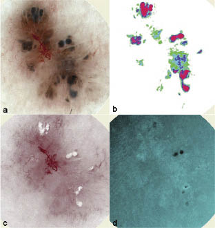
Fig 1. Pigmented basal cell carcinoma in dermoscopy view, (a) with large blue-grey ovoid nests, maple leaf-like areas, arborizing (tree-like) telangiectasia. In dermal melanin view (b) high concentration of dermal melanin (red) in ovoid nests. In these ovoid nests one can also see blood displacement (c) as white areas and collagen holes (d) as black areas.
Interesting comparisons can also be made regarding the localization of pigment in pigmented BCCs. According to Maloney et al. (1) dermal pigment is typically found at the tumour shoulders, but is occasionally located below the tumour, especially when melanocytes are found in deep tumour nodules. In our test set the DM is seen most commonly in the centre of the tumour, in large blue-grey ovoid nests and in blue-grey globules.
In this study we chose to focus exclusively on pigmented BCCs because one of the authors (KT, earlier Westerhoff) has previously carried out research into pigmented BCCs and dermoscopy (17) and we found it interesting to follow up with a study comparing dermoscopy and SIAscopy. Because of our findings that pigmented BCCs show the same SIAscopic features as previously presented for melanoma (18), further studies should be carried out comparing SIAscopic findings for pigmented BCCs and for melanomas. In this study we identified no SIAscopic features specific for pigmented BCCs, but it is possible that a larger study group may reveal patterns that might be of diagnostic use. Can it be a tool to estimate the depth of the pigmented BCC? Contradicting this is the fact that findings of dermal melanin can be seen in superficial BCCs (only two included in this study). Perhaps further studies on the blood images will lead to methods of showing the borders of the lesions, which would be a helpful tool when deciding on excision margins.
Further work should also be carried out to evaluate the correlation between SIAscopic findings and histopathology on tumours other than pigmented BCCs.
Our conclusion, in this limited setting, is that SIAscopy has no advantage over dermoscopy when diagnosing pigmented BCCs. We suggest that SIAscopy can be used as a complement to dermoscopy. If the examiner has no, or limited, knowledge of dermoscopy, SIAscopy can be misleading.
ACKNOWLEDGEMENT
We thank our colleague Dr Carin Sandberg for her contribution to the data collection.
Conflicts of interest: Clinical trials with Astron Clinica have been initiated. A-M. Wennberg has received fees for education of dermatologists.
REFERENCES
1. Maloney M, Jones D, Sexton F. Pigmented basal cell carcinoma; investigation of 70 cases. J Am Acad Dermatol 1992; 27: 74–78.
2. Betti R, Gualandri L, Cerri A, Inselvini E, Crosti C. Clinical features and histologic pattern analysis of pigmented basal cell carcinomas in an Italian population. J Dermatol 1997; 25: 691–694.
3. Bleehen S. Pigmented basal cell epithelioma. Br J Dermatol 1975; 93: 361–370.
4. Tezuka T, Ohkuma M, Hirose I. Melanosomes of pigmented basal cell epitheliomas. Dermatologica 1977; 154: 14–22.
5. Lao LM, Kumakiri M, Kiyohara T, Kuwahara H, Ueda K. Sub-populations of melanocytes in pigmented basal cell carcinoma: a quantitative, ultrastructual investigation. J Cutan Pathol 2001; 28: 34–43.
6. Krahn G, Gottlober P, Sander C, Peter RU. Dermatoscopy and high frequency sonography: two useful non-invasive methods to increase preoperative diagnostic accuracy in pigmented skin lesions. Pigment Cell Res 1998; 11: 151–154.
7. Steiner A, Pehamberger H, Wolff K. In vivo epiluminescence microscopy of pigmented skin lesions. II. Diagnosis of small pigmented skin lesions and early detection of malignant melanoma. J Am Acad Dermatol 1987; 17: 584–591.
8. Pehamberger H, Binder M, Steiner A, Wolff K. In vivo epiluminescence microscopy: improvement of early diagnosis of melanoma. J Invest Dermatol 1993; 100: S356–S362.
9. Steiner A, Pehamberger H, Binder M, Wolff K. Pigmented Spitz nevi: improvement of the diagnostic accuracy by epiluminescence microscopy. J Am Acad Dermatol 1992; 27: 697–701.
10. Binder M, Schwarz M, Winkler A, Steiner A, Kaider A, Wolff K, Pehamberger H. Epiluminescence microscopy: a useful tool for the diagnosis of pigmented skin lesions for formally trained dermatologists. Arch Dermatol 1995; 131: 286–291.
11. Nachbar F, Stolz W, Merkle T, Cognetta AB, Vogt T, Landthaler M, et al. The ABCD rule of dermatoscopy: high prospective value in the diagnosis of doubtful melanocytic skin lesions. J Am Acad Dermatol 1994; 30: 551–559.
12. Pazzini C, Pozzi M, Betti R, Vergani R, Crosti C. Improvement of diagnostic accuracy in the clinical diagnosis of pigmented skin lesions by epiluminescence microscopy. Skin Cancer 1996; 11: 159–161.
13. Binder M, Puespoeck-Schwarz M, Steiner A, Kittler H, Muellner M, Wolff K, Pehamberger H. Epiluminescence microscopy of small pigmented skin lesions: short-term formal training improves the diagnostic performance of dermatologists. J Am Acad Dermatol 1997; 36: 197–202.
14. Carli P, De Giorgi V, Naldi L, Dosi G. Reliability and inter-observer agreement of dermoscopic diagnosis of melanoma and melanocytic naevi. Eur J Cancer Prev 1998; 7: 397–402.
15. Stanganelli I, Serafini M, Cainelli T, Cristofolini M, Baldassari L, Staffa M, Bucchi L. Accuracy of epiluminescence microscopy among practical dermatologists: a study from the Emilia-Romagna region of Italy. Tumouri 1998; 84: 701–705.
16. Argenziano G, Soyer HP, Chimenti S, Talamini R, Corona R, Sera F, et al. Dermoscopy of pigmented skin lesions: results of a consensus meeting via the Internet. J Am Acad Dermatol 2003; 48: 679–693.
17. Menzies SW, Westerhoff K, Rabinovitz H, Kopf AW, McCarthy WH, Katz B. Surface microscopy of pigmented basal cell carcinoma. Arch Dermatol 2000; 136: 1012–1016.
18. Moncrieff M, Cotton S, Claridge E, Hall P. Spectrophotometric intracutaneous analysis: a new technique for imaging pigmented skin lesions. Br J Dermatol 2002; 146: 448–457.

