Akihiko Asahina1, Norihisa Ishii2, Hiromichi Kai1, Mizuho Yamamoto1 and Hideki Fujita1
1Department of Dermatology, Sagamihara National Hospital, 18-1 Sakuradai, Sagamihara, 228-8522 Kanagawa, and 2Department of Bioregulation, Leprosy Research Center, National Institute of Infectious Diseases, Tokyo, Japan. E-mail: asahina-tky@umin.ac.jp
Accepted March 31, 2007.
Sir,
Dowling-Degos disease (DDD) is a rare disorder characterized by acquired pigmented macules and papules in a reticulate pattern, particularly affecting the flexural areas and other major skin folds (1, 2). A female patient presented with dotted and reticulate pigmentation of the axilla, and was diagnosed as DDD both clinically and histopathologically. However, the asymmetrical distribution of the eruption was distinct from classical DDD. In contrast to recent reports in Caucasian patients with DDD (3, 4), we found no evidence of mutation of exon 1 of the keratin 5 (KRT5) gene, suggesting an aetiological heterogeneity of this disorder.
CASE REPORT
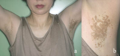
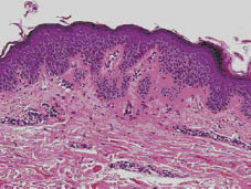
A 47-year-old Japanese woman presented at our department with development of reticulate pigmentation on her axilla. She had first noticed dotted pigmentation on her left axilla 8 years previously, which had gradually increased and coalesced. Similar pigmentation subsequently emerged on her right axilla 2 years previously. There were no clinical symptoms, such as pruritus, and she had no axillary hyperhidrosis. On examination, miliary brown macules and papules were seen distributed over the vault of her axilla, coalescing to give rise to reticulated patches. The lesion on the left side was more pronounced than that on the right (Fig. 1). The eruptions were slightly hyperkeratotic, with no apparent scaling on the surface. Some macules were in linear alignment, suggesting the presence of Koebner’s phenomenon. The patient was otherwise healthy. There was no pigmentation in other areas, such as the groin, neck or other flexures. No other family members had pigmentary abnormalities. A biopsy specimen from the early lesion on the right axilla showed slight orthokeratosis and acanthosis, characterized by irregular elongation of rete ridges and basal hyperpigmentation (Fig. 2). There was no papillomatosis. A subsequent biopsy from the older lesion on the left side showed an identical histological picture (not shown). Slight oedema and infiltration of lymphocytes around capillaries were seen in the upper dermis. Considering the characteristic clinical picture and the histopathological findings, a diagnosis of DDD was made. Repeated cryosurgery using liquid nitrogen was partially effective in resolving the lesion (not shown).
Fig. 1. (a) Multiple, small brown macules and papules in reticular distribution localized on the axilla. The lesion on the left side was more pronounced than that on the right. (b) Close-up of the left axilla.
Fig. 2. Histopathological picture showing irregular elongation of rete ridges and basal pigmentation (H&E stain ×200).
DISCUSSION
DDD (1, 2) usually presents in adults, commonly women, and most frequently during the fourth decade of life. Although DDD is usually inherited in an autosomal dominant fashion, some sporadic cases have been reported, as in our patient. The axilla and groin are the most common sites, but other areas may be involved, including the intergluteal and infra-mammary folds, neck, scalp, trunk and arms. Other possible cutaneous manifestations include perioral pitted scars and comedo-like lesions on the neck and back. Occasional cases have been reported with associated hidradenitis suppurativa, trichilemmal cysts, keratoacanthoma and squamous cell carcinoma. In addition, there are multiple reports of patients with the coexistence of DDD and reticulate acropigmentation of Kitamura. The histopathology of DDD typically shows filiform epithelial down-growth of epidermal rete ridges with basal hyperpigmentation (1, 2).
In our case, other cutaneous disorders that might primarily involve the axillary area, such as acanthosis nigricans, were unlikely, and the possibility of verruca plana or syringoma was also ruled out. Curiously, however, our case was unique and distinct from typical DDD, because the lesion was limited to the axilla, and the pigmentation initially developed on the left side, followed by the right, giving rise to a rather asymmetrical appearance. Considering that axillary pigmentation has developed consistently over time, other flexural areas may be similarly involved in the future (5). The asymmetrical distribution of pigmentation and likelihood of the coexistence of Koebner’s phenomenon indicate strongly that local stimuli may have triggered the onset and progression of the disease, although the lesion was asymptomatic and the patient did not remember scratching the axilla. Mechanical rubbing stimulation in daily life may be sufficient as a trigger. Contrary to the observation by Lee et al. (5) and Rubio et al. (6), the pigmentation was not preceded by erythema, suggestive of initial inflammation, and the biopsy specimen from both the early and old lesions did not reveal any clue to its pathogenesis.
Two recent reports have determined the location of a putative DDD gene by genome-wide linkage analysis. Li et al. (7) mapped this disorder to chromosome 17p13.3 in a Chinese family, and Betz et al. (3) to chromosome 12q in two German families. In the latter report, two loss-of-function mutations were identified in exon 1 of the KRT5 gene, leading to the truncation of KRT5 protein before the rod domain, in all affected family members and in six of an additional eight unrelated Caucasian patients with DDD. Liao et al. (4) also revealed a novel mutation in the same locus in the proband from a Spanish family with DDD. We therefore focused our genetic analysis on exon 1 of the KRT5 gene, and amplified a 667-bp DNA fragment spanning this region by polymerase chain reaction (PCR) with the designated primers and conditions (4)*; however, direct DNA sequencing of the PCR product did not reveal any mutation. While it remains possible that KRT5 gene mutation occurred elsewhere other than in exon 1, our results and those of other investigators (3, 7) indicate that DDD is a heterogeneous disease, and that patients with DDD do not necessarily have KRT5 haplo-insufficiency. Indeed, the reported clinical manifestations of DDD are diverse, and the racial differences may also be an important factor. Following this patient for future development of pigmentation, and additional reports of similar cases, may lead to further understanding of this rare disorder.
*The sequence of the primer K5EX1F was modified according to the consensus sequence of the KRT5 gene (AAAACC):
REFERENCES
1. Kim YC, Davis MD, Schanbacher CF, Su WP. Dowling-Degos disease (reticulate pigmented anomaly of the flexures): a clinical and histopathologic study of 6 cases. J Am Acad Dermatol 1999; 40: 462–467.
2. Li L, Nahm WK, Moskowitz P, Badiavas E, Danahy J. Brown macules symmetrically distributed on the neck, axillae, and thighs. Arch Dermatol 2003; 139: 657–662.
3. Betz RC, Planko L, Eigelshoven S, Hanneken S, Pasternack SM, Bussow H, et al. Loss-of-function mutations in the keratin 5 gene lead to Dowling-Degos disease. Am J Hum Genet 2006; 78: 510–519.
4. Liao H, Zhao Y, Baty DU, McGrath JA, Mellerio JE, McLean WH. A heterozygous frameshift mutation in the V1 domain of keratin 5 in a family with Dowling-Degos disease. J Invest Dermatol 2007; 127: 298–300.
5. Lee SJ, Lee HJ, Kim DW, Jun JB, Chung SL, Bae HI. A case of Dowling-Degos disease suggesting an evolutional sequence. J Dermatol 2000; 27: 591–597.
6. Rubio C, Mayor M, Martin MA, Gonzalez-Beato MJ, Contreras F, Casado M. Atypical presentation of Dowling-Degos disease. J Eur Acad Dermatol Venereol 2006; 20: 1162–1164.
7. Li CR, Xing QH, Li M, Qin W, Yue XZ, Zhang XJ, et al. A gene locus responsible for reticulate pigmented anomaly of the flexures maps to chromosome 17p13.3. J Invest Dermatol 2006; 126: 1297–1301.