Reine Moriuchi, Ken Arita*, Hideyuki Ujiie, Kazuo Kodama, Tadamichi Shimizu and Hiroshi Shimizu
Department of Dermatology, Hokkaido University Graduate School of Medicine, N15W7, Kita-ku, Sapporo 060-8638, Japan. *E-mail: ken_arita222@yahoo.co.jp
Accepted November 19, 2007.
Sir,
The non-tuberculous mycobacteria (NTM) are a heterogeneous group of acid-fast bacilli which differ from Mycobacterium tuberculosis. With the advent of the AIDS epidemic and the introduction of immunosuppressive therapies, the incidence of NTM-associated diseases has risen (1). In healthy individuals, cutaneous infection with NTM usually results in small abscess with sinus formation, whilst in immunocompromised hosts the abscess tend to be multiple and widespread (2, 3). We describe here an unusual case of a young healthy woman who developed multiple large subcutaneous abscesses caused by M. fortuitum.
CASE REPORT
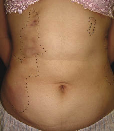
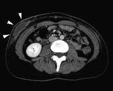
A 25-year-old Japanese woman presented with a 6-month history of painful indurated nodules on the abdomen. She was otherwise well with no history of general malaise or weight loss. Initially, the lesions appeared on her left abdominal wall and 3 months later, they had increased in size and were present bilaterally (Fig. 1). Cutaneous examination revealed multiple indurated nodules, the largest measuring 20 × 8 cm. She was afebrile with no lymphadenopathy, and no particular clinical sign of immunocompromised status was observed. Past medical and surgical history was unremarkable. White blood cell count was 6900 cells/mm3 and erythrocyte sedimentation rate was 8 mm/1st hour. Chest X-ray showed no abnormalities. Cushing’s syndrome was denied because of her normal body proportions, normal muscle strength, normotention, and good glucose tolerance. HIV testing was negative. A computed tomography scan revealed multiple subcutaneous abscesses on both sides of the abdominal wall (Fig. 2). The lesions were restricted to the subcutaneous area, with no involvement of the internal organs. A lesional skin biopsy including part of the abscess wall was taken. Histopathological findings showed diffuse necrosis and dense infiltration of neutrophils, lymphocytes and histiocytes in the lower dermis extending to the upper part of the subcutaneous fat. Multinuclear giant cells were also present. Incisional drainage from one of the abscesses was performed, resulting in the discharge of large amounts of yellow serous fluid. An acid-fast smear from this discharge was positive for mycobacteria. Microbiological culture on Ogawa culture medium produced milky-white colonies and DNA-based techniques revealed the presence of M. fortuitum. Subsequently, a diagnosis of cutaneous NTM infection was made and she started taking 2 oral antibiotics, levofloxacin (300 mg/day) and minocycline (200 mg/day) for 6 months. Six months later the lesions had completely regressed, leaving only slight subcutaneous indurations. To date, she remains healthy without any relapse.
Fig. 1. Clinical photograph on admission (6 months after the onset of the disease). Multiple subcutaneous abscesses on both sides of the abdomen (dotted lines). The largest was on the right side, measuring 20 × 8 cm.
Fig. 2. Computed tomography scan showing multiple subcutaneous abscesses containing large amounts of serous fluid (arrows). No other lesions were found.
Hendrick et al. (4) described a case of a 36-year-old woman who developed a large subcutaneous abscess on the left buttock. However, she had systemic lupus erythematosus for which she was taking oral prednisolone, which would have impaired her immune status. In contrast, our patient was in good health and the infection resulted in multiple, large abdominal abscesses, measuring up to 20 cm in diameter.
M. fortuitum is ubiquitous and has been found in soil and in various water resources, such as tap water (6) and ice-making machines (7). Iatrogenic procedures or trauma may result in cutaneous M. fortuitum inoculation in healthy subjects (5, 8–10), but a detailed inquiry into her medical history did not disclose any clues. This case report shows that M. fortuitum infection can result in large and multiple subcutaneous abscesses without obvious signs of immunocompromised status.
References
1. Wolinsky E. Mycobacterial disease other than tuberculosis. Clin Infect Dis 1992; 15: 1–12.
2. Murdoch ME, Leigh IM. Sporotrichoid spread of cutaneous Mycobacterium Chelonei. Clin Exp Dermatol 1989; 14: 309–312.
3. Wagner D, Young LS. Nontuberculous mycobacterial infections: a clinical review. Infection 2004; 32: 257–270.
4. Hendrick SJ, Jorizzo JL, Newton RC. Giant Mycobacterium fortuitum; abscess associated with systemic lupus erythematosus. Arch Dermatol 1986; 122: 695–697.
5. Muthusami JC, Vyas FL, Mukundan U, Jesudason MR, Govil S, Jesudason SR. Mycobacterium fortuitum; an iatrogenic cause of soft tissue infection in surgery. ANZ J Surg 2004; 74: 662–666.
6. Kauppinen J, Nousiainen T, Jantunen E, Mattila R, Katila ML. Hospital water supply as a source of disseminated Mycobacterium fortuitum infection in a leukemia patient. Infect Control Hosp Epidemiol 1999; 20: 343–345.
7. Labombardi VJ, O’brien AM, Kislak JW. Pseudo-outbreak of Mycobacterium fortuitum due to contaminated ice machines. Am J Infect Control 2002; 30: 184–186.
8. Sniezek PJ, Graham BS, Busch HB, Lederman ER, Lim ML, Poggemyer K, Rapidly growing mycobacterial infections after pedicures. Arch Dermatol 2003; 139: 629–634.
9. Subbarao EK, Tarpay MM, Marks MI. Soft-tissue infections caused by Mycobacterium fortuitum complex following penetrating injury. Am J Dis Child 1987; 141: 1018–1020.
10. Silvestre Salvador JF, Betlloch MI, Alfonso R, Ramon RL, Morell AM, Navas J. Disseminated skin infection due to Mycobacterium fortuitum in an immunocompetent patient. J Eur Acad Dermatol Venereol 1998; 11: 158–161.