The aim of this study was to test the hypothesis that particular clinical features of foreskin condylomata acuminata in Chinese male patients are associated with diabetes. A prospective study enrolled 126 men presenting with foreskin condylomata acuminata from 2001 to 2006. Mean age was 46 years (age range 25–74 years) and mean duration of disease was 4.8 months (range 1–18 months). Patients were divided into two groups according to clinical features. In group 1, 42 men had distinctive signs such as redundant prepuce, crown warts circling the entire preputial ring, maceration, fissures, phimosis and balanitis, and 37 of 42 (88%) patients were found to have concurrent type 2 diabetes, furthermore 32 of these 37 patients had an insidious onset and were previously undiagnosed. In group 2, 84 male patients did not have those distinctive clinical features and type 2 diabetes was found in only 10 cases (11.9%, p < 0.0001, Fisher’s exact test). These clinical features strongly suggest the presence of diabetes. Therapy should address diabetes and condylomata concurrently. Key words: condylomata acuminata; diabetes mellitus; clinical features.
(Accepted May 20, 2008.)
Acta Derm Venereol 2008; 88: 578–583.
Xiu L. Wang, Department of Dermatology and Venereology, Shanghai Skin Diseases and STD Hospital, Wuyi Rood 196, Shanghai 200050, P. R. China. E-mail: xiuliwang2001@yahoo.com.cn
Sexually transmitted infections (STI) in China began to increase in the early 1980s after the “opening up” of the country to outside influences. Among STI the incidence of condylomata acuminata in particular has increased during the past two decades (1). Genital condylomata acuminata is the most common STI in China and is caused by human papillomavirus (HPV) (2). In men, warts can occur anywhere on the penis, although the coronal sulcus is the most common site. Their color is generally gray, pale yellow or pink. Treatment of condylomata acuminata may be difficult, and a wide range of therapeutic procedures, such as cryotherapy, surgery, laser, keratolytic agents, cytostatic agents and immunologic therapy, have been applied.
Diabetes mellitus (DM) is rapidly reaching pandemic proportions in many parts of the world, largely owing to an increase in type 2 DM. The incidence of DM in China has been increasing during recent years, as lifespan is extended, living standards are elevated and “westernized” lifestyles are adopted (3). A common finding in DM is that the skin and mucosa are found to be colonized (and can be infected) by pathogenic microorganisms, such as bacteria, viruses and fungi (e.g. Candida albicans) (4).
In the years prior to 2001, we noted some distinctive clinical features of condylomata acuminata in more than 12 male patients at the STI outpatient clinic of Shanghai Skin Diseases and STD Hospital. The patients presented with very distinctive but similar clinical manifestations, such as a large exophytic growth of rooster comb warts completely circling the preputial ring, redundant prepuce, phimosis, maceration and fissures, plus secondary balanitis. The hypothesis was proposed that these particular clinical features were associated with diabetes (whether diagnosed or undiagnosed).
To confirm the hypothesis and explore suitable treatment approaches, a prospective study was carried out, which enrolled 126 Chinese male patients presenting with genital condylomata acuminata during 2001 to 2006. Fasting plasma glucose was measured and treatment was designed to address the DM as well as the warts if DM was confirmed. The relationship between the clinical features of condylomata acuminata and DM was also analyzed.
PATIENTS AND METHODS
A total of 133 consecutive male patients with genital condylomata acuminata without anal lesions were enrolled by one clinic at the Shanghai Skin Diseases and STD Hospital, China, between 2001 and 2006 after receiving written informed consent. The study was approved by the hospital research committee. The diagnosis of condylomata acuminata was made by clinical and histopathological examination. Gonococcal and non-gonococcal urethritis and HIV-seropositive patients were excluded from the enrollment. In this study, 5 patients were excluded because of coexisting gonococcal or non-gonococcal urethritis. Study parameters included age, a complete history, clinical presentation and physical findings, presence of previous or coexisting STI, mode of treatment, residual and recurrent disease. Rapid plasma regain (RPR) and Treponema pallidum haemagglutination (TPHA) testing for syphilis, and fasting plasma glucose testing for DM were performed. The diagnosis of DM was made according to the American Diabetes Association (ADA) Expert Committee that updated the diagnostic glucose levels for DM (i.e. ≥ 126 mg/dl) in 1997 (5).
The patients were divided into two groups according to clinical features. In group 1, patients (n = 42) presented with distinctive clinical features, such as redundant prepuce, crown warts circling preputial ring, maceration, fissures, phimosis and balanitis (Table I). In group 2, patients (n = 84) did not have these special features, and warts were found to be randomly located anywhere on the penis. Two borderline cases that could not be clearly assigned to group 1 or 2 were excluded.
Table I. Clinical data of group 1
|
Patient no.
|
Age (years)
|
Duration of warts (months)
|
History of diabetes (months)
|
Previous treatment of warts
|
Plasma glucose (mg/dl)a
|
Clinical appearance
|
HPV
|
Balanitis
|
|
1
|
32
|
4
|
0
|
No
|
187
|
Prepuce, phimosis
|
Low-risk
|
Yes
|
|
2
|
76
|
6
|
0
|
Salicylic acid
|
183
|
Prepuce, phimosis
|
Low-risk
|
Yes
|
|
3
|
25
|
3
|
0
|
Laser
|
224
|
Prepuce, frenulum, glans, phimosis
|
Low-risk
|
Yes
|
|
4
|
54
|
1
|
0
|
No
|
185
|
Prepuce, frenulum
|
Low-risk
|
No
|
|
5
|
50
|
1
|
0
|
No
|
169
|
Prepuce
|
High-risk
|
Yes
|
|
6
|
54
|
4
|
0
|
Salicylic acid
|
130
|
Prepuce
|
Low-risk
|
No
|
|
7
|
51
|
6
|
0
|
Laser, salicylic acid
|
127
|
Prepuce, frenulum, phimosis
|
Low-risk
|
Yes
|
|
8
|
54
|
8
|
0
|
Laser
|
288
|
Prepuce, paraphimosis
|
Low-risk
|
Yes
|
|
9
|
54
|
8
|
0
|
Laser,
|
131
|
Prepuce, paraphimosis
|
Low-risk
|
Yes
|
|
10
|
74
|
12
|
0
|
Podophyllin
|
129
|
Prepuce, frenulum, phimosis
|
Low-risk
|
Yes
|
|
11
|
50
|
5
|
0
|
Podophyllin
|
233
|
Prepuce, frenulum, phimosis
|
Low-risk
|
No
|
|
12
|
36
|
4
|
0
|
Laser
|
197
|
Prepuce, frenulum, phimosis
|
Low-risk
|
Yes
|
|
13
|
36
|
5
|
0
|
Laser, podophyllin
|
172
|
Prepuce, frenulum, glans, phimosis
|
Low-risk
|
Yes
|
|
14
|
35
|
8
|
0
|
Laser, salicylic acid
|
128
|
Prepuce, other area
|
Low-risk
|
Yes
|
|
15
|
55
|
6
|
0
|
Salicylic acid
|
129
|
Prepuce
|
Low-risk
|
No
|
|
16
|
37
|
6
|
0
|
Salicylic acid
|
210
|
Prepuce, frenulum, phimosis
|
Low-risk
|
Yes
|
|
17
|
50
|
2
|
34
|
No
|
140
|
Prepuce, phimosis
|
Low-risk
|
No
|
|
18
|
67
|
2
|
0
|
No
|
227
|
Prepuce, coronal sulcus, glans, other area
|
Low-risk
|
Yes
|
|
19
|
30
|
5
|
0
|
No
|
198
|
Prepuce, phimosis
|
Low- and high-risk
|
Yes
|
|
20
|
43
|
6
|
6
|
Laser
|
193
|
Prepuce, coronal sulcus, other
|
Low-risk
|
Yes
|
|
21
|
50
|
4
|
0
|
Laser
|
126
|
Prepuce, glans, paraphimosis
|
Low- and high-risk
|
Yes
|
|
22
|
43
|
1
|
0
|
Podophyllin
|
246
|
Prepuce, frenulum
|
Low-risk
|
Yes
|
|
23
|
34
|
6
|
0
|
Laser, salicylic acid
|
217
|
Prepuce
|
Low-risk
|
No
|
|
24
|
46
|
4
|
36
|
Laser, podophyllin
|
160
|
Prepuce, frenulum, phimosis
|
Low-risk
|
Yes
|
|
25
|
36
|
2
|
0
|
Laser, salicylic acid
|
142
|
Prepuce, glans
|
Low-risk
|
Yes
|
|
26
|
51
|
4
|
0
|
Laser
|
209
|
Prepuce, glans
|
High-risk
|
Yes
|
|
27
|
47
|
4
|
0
|
Laser, salicylic acid
|
126
|
Prepuce, frenulum, glans, paraphimosis
|
Low-risk
|
Yes
|
|
28
|
58
|
18
|
0
|
Laser
|
92
|
Prepuce, other, phimosis
|
High-risk
|
Yes
|
|
29
|
35
|
5
|
0
|
Laser
|
91
|
Prepuce, other
|
Low-risk
|
No
|
|
30
|
34
|
2
|
0
|
Podophyllin
|
136
|
Prepuce
|
High-risk
|
Yes
|
|
31
|
48
|
1
|
0
|
Laser
|
88
|
Prepuce
|
Low- and high-risk
|
No
|
|
32
|
55
|
4
|
0
|
No
|
82
|
Prepuce, phimosis
|
Low-risk
|
No
|
|
33
|
48
|
2
|
0
|
Laser
|
129
|
Prepuce, phimosis
|
Low-risk
|
Yes
|
|
34
|
33
|
3
|
0
|
Podophyllin
|
213
|
Prepuce, frenulum, glans, phimosis
|
Low-risk
|
Yes
|
|
35
|
56
|
1
|
0
|
No
|
187
|
Prepuce, frenulum
|
Low-risk
|
No
|
|
36
|
49
|
1
|
0
|
No
|
165
|
Prepuce
|
Low- and high-risk
|
Yes
|
|
37
|
52
|
4
|
0
|
Imiquimod
|
138
|
Prepuce
|
Low-risk
|
No
|
|
38
|
61
|
6
|
0
|
Laser, salicylic acid
|
129
|
Prepuce, frenulum, phimosis
|
Low-risk
|
Yes
|
|
39
|
35
|
8
|
0
|
Laser
|
285
|
Prepuce, paraphimosis
|
Low-risk
|
Yes
|
|
40
|
43
|
8
|
24
|
Laser, imiquimod
|
130
|
Prepuce, paraphimosis
|
Low-risk
|
Yes
|
|
41
|
49
|
12
|
0
|
Imiquimod
|
124
|
Prepuce, frenulum, phimosis
|
Low-risk
|
No
|
|
42
|
57
|
5
|
0
|
Podophyllin
|
237
|
Prepuce, frenulum, phimosis
|
Low-risk
|
Yes
|
aNormal range < 126 mg/dl. HPV: human papillomavirus.
Specimens were tested for HPV DNA by the US Food and Drug Administration (FDA)-approved Hybrid Capture 2 HPV DNA assay (Digene, Silver Spring, MD, USA) and analyzed for the presence of low-risk HPV types 6, 11, 42, 43 and 44 and high-risk HPV types 16, 18, 31, 33, 35, 39 45, 51, 52, 56, 58, 59 and 68. The hybrid capture system is a capture molecular hybridization assay that uses chemiluminescence detection in microtiter plates. Samples were classified positive for HPV DNA if the relative light-unit reading obtained from the luminometer was equal to or greater than the mean of positive control values according to the manufacturer’s instructions.
The first follow-up visit was carried out one month after condylomata acuminata treatment in all cases. Thereafter, clinical follow-ups were scheduled at 2, 4, 6 and 12 months. The following data were collected: demographic anamnestic characteristics, size and location of the lesions, type of treatment, plasma glucose level and signs of neoplasia. The female partners of high-risk HPV patients were also examined for HPV DNA types and signs of neoplasia.
Fisher’s exact test was used for statistical analysis.
RESULTS
History
In group 1, among 42 patients with the distinctive clinical features of condylomata acuminata, only 5 of 42 cases (11.9%) had been previously diagnosed with DM. The other 37 (88.1%) had no history of DM before attending our hospital. Thirty-three of 42 (78.6%) patients had received prior treatment for condylomata acuminata. Among 33 patients with a history of treatment, carbon dioxide (CO2) laser was applied in 21 (63.6%), salicylic acid, imiquimod, podophyllin were used in 12 (36.4%) men (Table I). Stenosis of the preputial ring was found in 15 of 21 (71.4%) patients after treatment with CO2 laser.
Among the 84 patients in group 2 without distinctive clinical features, 11.9% (10/84) of cases had DM, 72.6% (61/84) had prior treatment for penile warts, and 67.2% (41/61) had received CO2 laser therapy. There was no significant difference in the use of CO2 laser between the two groups of patients. However, the occurrence of stenosis of the preputial ring was only 2.4% (1/41) in group 2 and was much lower than that in group 1 (p < 0.0001) (Table II).
Clinical manifestation
In group 1, the mean age of the participants was 47 years (age range 25–74 years). All patients included in the study had had condylomata acuminata for 1–18 months. The mean duration was 4.9 months. All the patients presented with similar clinical features (Fig. 1A–C). They are described as follows:
• All of the patients had redundant prepuce (the glans was covered completely by the prepuce).
• The warts presented as exophytic growths, with rooster comb appearance and completely circled the preputial ring in all cases. Maceration, fissures and bleeding could often be observed on the prepuce (Fig. 1C). Phimosis and/or paraphimosis were often present (Fig. 1B). The frenulum prepuce could also be covered with warts. The coronal sulcus and glans were seldom involved. However, no warts were found on the scrotum or base of the penis.
• Balanitis was commonly seen, especially as candidal balanitis. In group 1, balanitis was found in 30 of 42 (71.4%) patients, and of these 25 of 30 (83.3%) cases were candidal balanitis as confirmed by fungal culture.
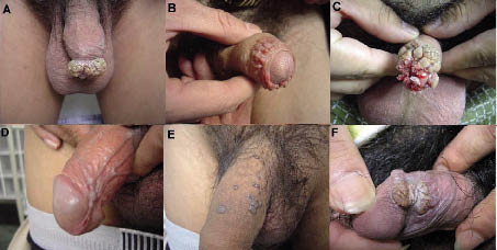
Fig. 1. (A) Group 1 patient 13 with condylomata acuminata for 5 months. The warts presented as exophytic growths circling the preputial ring. Phimosis and candidal balanitis was present. (B) Group 1 patient 4 with condylomata acuminata for 1 month. The warts circled the preputial ring and phimosis can be seen. (C) Group 1 patient 19 with condylomata acuminata for 5 months. The warts presented as exophytic growths circling the preputial ring. Phimosis was present. Maceration, fissures and bleeding on the prepuce and candidal balanitis could be observed. (D) Group 2 patient with condylomata acuminata for 2 months. The warts were located only on the frenulum. (E) Group 2 patient with condylomata acuminata for 8 months. The warts were randomly distributed on the penis. (F) Group 2 patient with condylomata acuminata for 4 months. One large wart covered the coronal sulcus.
In group 2, the mean age of the participants was 46 years (age range 21–73 years). The mean duration of genital condylomata acuminata was 4.4 months (range 1–15 months). The warts were randomly distributed on the penis (Fig. 1D–F). More than half (43/84) of the patients had warts on the coronal sulcus and prepuce (Fig. 1F). Redundant prepuce was found in only 29.7% (25/84 men). The rate was much lower than that in group 1 (p < 0.0001). The distinctive clinical features found in group 1 were absent from this group.
Laboratory examination
Among 42 patients of group 1, 9.5% (4/42) of the men were RPR positive (serum titer > 1:8) and TPHA positive (serum titer > 1:80), 4 of 42 (9.5%) patients harbored high-risk HPV, 4 of 42 (9.5%) harbored both high- and low-risk HPV, 34 of 42 (81%) cases harbored only low-risk HPV. There was no significant difference among these measurements between groups 1 and 2 (Table II).
Diabetes mellitus
All patients were tested for fasting plasma glucose. In group 1, excluding 5 patients with previous history of DM, 32 cases were found with plasma glucose levels equal or higher than 126 mg/dl. Overall, 37 of 42 patients (88.1%) were diagnosed as having type 2 DM according to the ADA Expert Committee values (4). Only 11.9% (5/42 patients) were found to have normal levels of fasting plasma glucose. In group 2, only 11.9% (10/84 patients) had plasma glucose levels consistent with type 2 DM (p < 0.0001 compared with group 1).
Among the patients with DM in group 1, 56.8% (21/37 patients) had a positive family history of DM, 59.5% (22/37 patients) were overweight (body mass index, BMI > 25 kg/m2) or obese (BMI > 30 kg/m2), and a history of high-fat diet and a sedentary lifestyle was found in 51.4% (19/37 patients). Two of 37 cases (5.4%) were under 30 years of age, 22 of 37 (59.5%) were 31–50 years of age, the other 13 of 37 patients (35.1%) were over 51 years of age, i.e. almost two-thirds of patients were younger than 50 years of age.
Balanitis and diabetes mellitus
Among 37 cases with DM in group 1, 30 patients (81.1%) had balanitis and, of these, 83.3% (25/30 patients) were candidal balanitis. However, among 5 of 42 patients without DM, only one case (20%) was observed with balanitis. No candidal infection was found. The incidence of balanitis was much higher in the patients with DM than in those without DM (p < 0.0001). In group 2, balanitis was observed in only 3.6% (3/84) patients (p < 0.0001 compared with group 1) (Table II). Only one of these 3 patients had DM.
Table II. Statistical comparison (Fisher’s exact test) of clinical data in groups 1 and 2
|
|
Cases n
|
Distinctive clinical features n (%)
|
Type 2 diabetes n (%)
|
Stenosis of preputial ring n (%)
|
Balanitis n (%)
|
Redundant prepuce n (%)
|
Recurrence n (%)
|
High-risk HPV n (%)
|
RPR and TPHA positive n (%)
|
|
Group 1
|
42
|
42 (100)
|
37 (88.1)
|
15/21 (71.4)
|
31 (71.4)
|
42 (100)
|
13 (31)
|
8 (19)
|
4 (9.5)
|
|
Group 2
|
84
|
0 (0)
|
10 (11.9)
|
1/41 (2.4)
|
3 (3.6)
|
25 (29.7)
|
27 (32.1)
|
17 (20.2)
|
9 (10.7)
|
|
p-value
|
|
0.000
|
0.000
|
0.000
|
0.000
|
0.000
|
0.892
|
0.874
|
0.836
|
HPV: human papillomavirus; RPR: rapid plasma regain; TPHA: Treponema pallidum haemagglutination.
Treatment
In the cases of condylomata acuminata associated with DM, management of DM was carried out during the same therapy session. After the patient’s plasma glucose was under control (e.g. < 110 mg/dl), patients were advised to undergo treatment for condylomata acuminata.
Of 37 patients with DM in group 1, 11 cases (29.7%) with plasma glucose over 200 mg/dl were treated with insulin, either alone or in combination with oral agents, such as sulfonylureas and/or biguanides. Diet and exercise alone were successfully used as therapy in 13 patients (35.1%). The other 13 cases (35.2%) required sulfonylureas and/or biguanides in addition to diet and exercise.
In group 1, because all patients presented with redundant prepuce and most of the warts circled the prepuce, more than one-third of the patients presented with stenosis of preputial ring consequent upon previous CO2 laser therapy. Therefore, circumcision was generally performed on these patients. After surgery, CO2 laser was employed in cases with warts on the frenulum prepuce and/or glans. The management in group 2 was similar to that in group 1 without the need for circumcision.
Terbinafine cream was applied for treatment of the patients with candidal balanitis. Mupirocin ointment was employed for treatment of other patients with non-candidal balanitis.
Follow-up
Follow-up of patients treated by circumcision and/or CO2 laser ranged from 6 to 12 months. The warts recurred in 31% (13/42 cases), 1–3 months after the treatment in patients in group 1. A recurrence rate of 32.1% (27/84 patients) was found in group 2. There was no significant difference between recurrence rates in the two groups. All 25 patients of high-risk HPV positive became high-risk HPV negative during follow-up. Their female partners were also examined during follow-up. Seven were lost during follow-up and 11 out of 18 (61.1%) were high-risk HPV-positive and 3 developed cervical intraepithelial neoplasia lesions.
DISCUSSION
The incidence of DM in China has been increasing, following recent economic development and the changing lifestyle over the last two decades, for instance high-fat diets, less labor-intensive employment and lower levels of physical exercise (3). It has been estimated that by 2010 there will be more than 30 million Chinese patients with DM (6). A family history of DM (7), being overweight (8) and a sedentary lifestyle (9) have been recognized for centuries as risk factors.
In group 1 of this study, 64.9% patients with DM were younger than 50 years. Except for five cases (13.5%) diagnosed when seeking the treatment of condylomata acuminata, 32 of 37 (86.5%) cases showed no history of DM. We found that most of the patients presented with insidious onset and were unrecognized before being diagnosed with condylomata acuminata. In addition, the risk factors for DM, such as positive family history, overweight, obesity and sedentary lifestyle, were found in more than half of the patients, which agreed with previous studies.
In China most men are uncircumcised. The presence of the foreskin, especially a redundant prepuce, appears to be an important risk factor for penile intraepithelial neoplasia and squamous cell carcinoma (10). The foreskin also could provide a suitable site for infection with HPV, C. albicans and bacteria (11).
In group 1 of the present study, 42 patients with foreskin condylomata acuminata presented distinctive clinical features, such as redundant prepuce, crown warts circling the preputial ring, maceration, fissures, phimosis and balanitis. Remarkably, 37 of 42 (88.1%) cases were found to have concurrent type 2 DM. In contrast, in group 2 these distinctive clinical features were absent and type 2 DM was rare. These findings suggested that the observation of distinctive clinical features of condylomata acuminata strongly suggests the presence of DM. The confirmation and treatment of DM should be strongly recommended.
Candidal balanitis was a common complication found in this study. All of the 25 cases with candidal balanitis were confirmed in patients with DM. The findings suggest that elevated plasma and urine glucose, together with a redundant prepuce provides a suitable environment for C. albicans infection. It is known that the coronal sulcus is usually the most common site of warts. However, the coronal sulcus, interior of the prepuce and the glans were seldom involved in this study. It is possible that this observation could be explained by HPV infection being somehow inhibited by existing C. albicans infection.
Although it is well known that DM is a predisposing factor for infection in general, and for cutaneous infections in particular, the mechanisms for this predisposition are not completely understood (12). Polymorphonuclear leukocyte adherence, chemotaxis, and phagocytosis are reduced (13), and bactericidal activity may also be impaired (14). Cutaneous responses to antigen challenges and measures of T-cell function may be depressed (15). We have not been able to trace any previous reports that DM increases the incidence or severity of HPV infection. One report found no difference in genital HPV amongst pregnant diabetic women compared with normal pregnant women (16).
Unsatisfactory previous treatment was observed in 33 patients and 15 of 21 patients presented with stenosis of the preputial ring after previous CO2 laser therapy. These findings suggest that conventional treatments may not be very effective for patients with concurrent DM. Our preferred therapeutic approach is, firstly, to initiate proper and effective treatment of DM, secondly, to perform circumcision as early as possible in order to remove most of the warts; and, thirdly, to eradicate the remaining warts on the frenulum prepuce and glans with CO2 laser. It should be emphasized, however, that wide laser ablation should be avoided because of the severe sequela of stenosis of the preputial ring.
DM is occurring with increasing frequency in the world, and the rate of increase is especially high in some developing countries. It is likely that the incidence of condyloma associated with DM is increasing at the same time. Awareness of this should prompt the clinician carefully to measure and control the plasma glucose level when a distinctive type of male foreskin condylomata acuminata is seen. Our study strongly suggests a combined approach of treating both diseases simultaneously.
REFERENCES
1. Shao C, Liang G. STD epidemiologic analysis at national surveillance spots in the period 1987–1990. Chin Med Sci J 1992; 7: 40–43.
2. Dupin N. Genital warts. Clin Dermatol 2004; 22: 481–486.
3. Wong KC, Wang Z. Prevalence of type 2 diabetes mellitus of Chinese populations in Mainland China, Hong Kong, and Taiwan. Diabetes Res Clin Pract 2006; 73: 126–134.
4. Odom RB. Skin and soft tissue infections in special populations. Cutis 2004; 73: 26–29.
5. Genuth S, Alberti KG, Bennett P, Buse J, Defronzo R, Kahn R, et al. Follow-up report on the diagnosis of diabetes mellitus. Diabetes Care 2003; 26: 3160–3167.
6. Amos AF, McCarty DJ, Zimmet P. The rising global burden of diabetes and its complications: estimates and projections to the year 2010. Diabet Med 1997; 14 Suppl 5: S1–S85.
7. Libman IM, Arslanian SA. Prevention and treatment of type 2 diabetes in youth. Horm Res 2007; 67: 22–34.
8. Ford ES, Williamson DF, Liu S. Weight change and diabetes incidence: findings from a national cohort of US adults. Am J Epidemiol 1997; 146: 214–222.
9. Helmrich SP, Ragland DR, Leung RW, Paffenbarger RS, Jr. Physical activity and reduced occurrence of non-insulin-dependent diabetes mellitus. N Engl J Med 1991; 325: 147–152.
10. Velazquez EF, Bock A, Soskin A, Codas R, Arbo M, Cubilla AL. Preputial variability and preferential association of long phimotic foreskins with penile cancer: an anatomic comparative study of types of foreskin in a general population and cancer patients. Am J Surg Pathol 2003; 27: 994–998.
11. Porter WM, Francis N, Hawkins D, Dinneen M, Bunker CB. Penile intraepithelial neoplasia: clinical spectrum and treatment of 35 cases. Br J Dermatol 2002; 147: 1159–1165.
12. Joshi N, Caputo GM, Weitekamp MR, Karchmer AW. Infections in patients with diabetes mellitus. N Engl J Med 1999; 341: 1906–1912.
13. Delamaire M, Maugendre D, Moreno M, Le Goff MC, Allannic H, Genetet B. Impaired leucocyte functions in diabetic patients. Diabet Med 1997; 14: 29–34.
14. Gallacher SJ, Thomson G, Fraser WD, Fisher BM, Gemmell CG, MacCuish AC. Neutrophil bactericidal function in diabetes mellitus: evidence for association with blood glucose control. Diabet Med 1995; 12: 916–920.
15. Saiki O, Negoro S, Tsuyuguchi I, Yamamura Y. Depressed immunological defence mechanisms in mice with experimentally induced diabetes. Infect Immun 1980; 28: 127–131.
16. Hietanen S, Ekblad U, Pelliniemi TT, Syrjanen K, Helenius H, Syrjanen S. Type I diabetic pregnancy and subclinical human papillomavirus infection. Clin Infect Dis 1997; 24: 153–156.


