Lotus Mallbris1, Fredrik von Bergen2, Marianne van Hage3 and Mona Ståhle1
1Dermatology and Venereology Unit, 3Clinical Immunology and Allergy Unit, Department of Medicine Solna, Karolinska Institutet, and 2Department of Radiology, Karolinska University Hospital, SE-171 76, Stockholm, Sweden. E-mail: lotus.mallbris@ki.se
Accepted June 2, 2009.
Sir,
Efalizumab is a recombinant humanized monoclonal IgG1 antibody directed against CD11a, leading to blockage of lymphocyte function-associated antigen (LFA)-1-mediated T-cell adhesion and subsequent inhibition of T-cell activation and trafficking to sites of cutaneous inflammation (1).
Efalizumab was approved in 2004 for the treatment of moderate–severe psoriasis in patients not responding to, or intolerant to, traditional systemic antipsoriatic therapy, and was administered as a subcutaneous injection once weekly.
Until now, global exposure of patients to efalizumab was estimated at 47,000 patient-years (2). However, efalizumab was recently withdrawn from the European, Canadian and US markets due to a potential risk for developing progressive multifocal leukoencephalopathy (PML), a rare, demyelinating disease of the central nervous system caused by an opportunistic viral infection and usually leading to death or severe disability (2).
The most common adverse reactions associated with efalizumab include headache, chills, fever, nausea and myalgia, developing within 2 days of the first two injections. Hypersensitivity reactions, including dyspnoea, asthma, urticaria, angioedema and maculopapular rash, have also been reported (3).
CASE REPORT
A 63-year-old man presented with severe plaque psoriasis since 15 years, which had been treated previously with methotrexate and narrowband ultraviolet-B, both of which were ineffective.
The patient had no other medical conditions and no prior history of allergies. At the initial visit, complete blood cell count, liver and renal function tests, urine analysis and tests for hepatitis revealed no abnormalities. His Psoriasis Area and Severity Index (PASI) score was 15 without arthritis (4).
Efalizumab treatment was started, at an initial dose of 0.7 mg/kg subcutaneously, followed by 1 mg/kg/week (1). Regular clinical and laboratory examinations were performed. The patient achieved PASI-90 after 6 weeks and almost complete clearance of psoriasis after 12 weeks.
At week 15 of efalizumab treatment he began to feel a tingling sensation in the gingiva, which over 2 days progressed to a swelling of the gingiva, tongue and lips. All symptoms resolved spontaneously after 3 days.
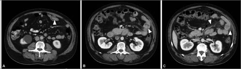
One week later, 3 days after taking the next injection of efalizumab, he was admitted to the emergency department with swelling of the tongue. He presented with angioedema with swelling of the periorbital area, cheek, tongue and lips, but without respiratory distress. His vital signs were stable. A few hours later, the angioedema worsened, involving his torso and arms, and he developed acute abdominal pain. Laboratory tests showed normal liver function, lipase and C-reactive protein. Serum amylase was slightly elevated (3.57 µ/l; normal: 0.40–2.00) and leucocyte count was 12.5 × 10⁹/l (normal: 3.5–8.8 × 10⁹/l). An abdominal computerized tomography (CT) scan showed an invagination in the distal jejunum (5) (Fig. 1A). After 3 days, his symptoms resolved spontaneously and he was discharged.
Fig. 1. Abdominal computerized tomography (CT) scan. (A) The first CT scan, showing an invagination in the distal jejunum (arrowhead). (B) A second CT scan after a new bout of abdominal pain, showing an invagination in the proximal jejunum (arrowhead). (C) Abdominal fluid in proximity to the invagination (arrowhead 1), along with general induration of the mesentery (arrowhead 2).
Following the next dose of efalizumab, the patient developed a new episode of swelling of the face, neck and extremities as well as acute abdominal pain. He was readmitted and explorative surgery was considered. Laboratory tests showed elevated serum amylase 4.62 µcat/l (0.40–2.00) and a high leucocyte count 16.6 × 10⁹/l. C4 was 0.42 g/l (0.12–0.31), while C1-esterase-inhibitor assay was normal. A new abdominal CT scan showed an invagination in the proximal jejunum (Fig. 1B) and abdominal fluid in proximity to the invagination, along with general induration of the mesentery (5) (Fig. 1C). Small bowel angioedema was diagnosed, and efalizumab treatment was discontinued. The swelling and abdominal pain resolved gradually and disappeared within 3 weeks. No further episodes of angioedema recurred in the ensuing 3 months. It is noteworthy that his psoriasis remained stable with no flare-up for 3 months.
DISCUSSION
Although the possibility of an unrelated cause cannot be excluded, in our opinion, the angioedema, in this case, was probably mediated by efalizumab. There are several ways in which efalizumab may have precipitated the angioedema: by interacting with IgE antibody on mast cells/basophils; by IgE-independent mast cell release; or by inappropriate activation of the complement cascade (6).
When analysing the presumed IgE-sensitivity to efalizumab, by incubating the patient’s basophils with serial dilutions of efalizumab (0.1–10000 μg/ml), we found no upregulation of CD63 and CD203c by flow cytometry. Thus, it is unlikely that the reaction was mediated by IgE (7).
REFERENCES