Georgios Gaitanis, Georgia Mitsou, Georgia Tsiouri, Ioannis Alexis and Ioannis D. Bassukas
Department of Skin and Venereal Diseases, University of Ioannina Medical School, GR-45110 Ioannina, Greece. E-mail: ibassuka@cc.uoi.gr
Accepted March 17, 2010.
Beyond surgical excision, curettage, cautery, cryosurgery, radiotherapy, laser, and photodynamic therapy have been advocated for the treatment of Bowen’s disease, an in situ skin carcinoma that has a 3–5% risk of progression to invasive neoplasm (1, 2). Selection criteria for the different modalities include the size and/or number of the lesions (1–3), localization of the lesion in poor wound-healing regions, such as the legs (3–5), and/or limitations in the availability of certain modalities. There is heterogeneity in the various therapeutic outcomes with cure rates ranging from 47–94% (1, 2, 6–9). In this study, prompted by the need for alternative treatments of Bowen’s disease, we evaluated ”immunocryosurgery”, i.e. a combination modality of cryosurgery during continuing topical imiquimod (10) which has been used successfully to treat basal cell carcinoma and lentigo maligna (11,12).
MATERIALS AND METHODS
Eight patients (5 women and 3 men; age range 52–87 years, mean 71.9 years) with 11 biopsy-proven Bowen’s disease lesions (Table I) were recruited between January and December 2007. The study was approved by the local hospital ethics committee according to the principles of the Declaration of Helsinki, and all patients provided informed consent. Treatment consisted of daily application of 5% imiquimod cream (Aldara®; Meda, Athens, Greece) to the lesion and a 5 mm rim of surrounding skin for a total of 5–9 weeks (Table I). At the clinic visit 2–3 weeks after starting imiquimod treatment liquid nitrogen cryosurgery was applied (open spray; two freeze-thaw cycles, 10–20 s freezing time each) to the inflamed skin including a 5-mm rim around the clinical margins of the lesions (Table I). Lesions of area > 1.5 cm2 were treated in approximately 1 cm2 subsections. Patients were instructed to continue imiquimod after cryosurgery without interruption and were evaluated every 3 weeks until the end of treatment. Persistence of erosion 3 weeks after ceasing topical imiquimod was considered as indicative of residual tumour (= partial response) and treatment with an additional cryosurgery course and/or extension of the application of imiquimod for a further 3 weeks was decided (Table I). Fusidic acid 2% cream (Fucidin®, Leo Pharma, Athens, Greece) 2× daily for 1 week and strict sun protection were prescribed after discontinuation of imiquimod. The follow-up period was calculated from cessation of imiquimod application (= endpoint of treatment) and visits were scheduled at 1 month, every 3 months for the following year and at 6-month intervals thereafter (up to 30 June 2009).
Table I. Treatment of Bowen’s disease with immunocryosurgery. Patients’ demographic data, core tumour characteristics, treatment parameters and outcome. No patients experienced any relapses
| Patient/age/sex | Tumour | Anatomical localization (side)a | Size (mm) | Precryosurgery imiquimod (weeks) | Cryosurgery: number of freeze-thaw cycles × freezing time (s) | Post-cryosurgery imiquimod (weeks) | Follow-up (months) |
| 1/72/F | 1 | Mandible (R) | 21 × 18 | 3 | 2 × 20 | 6b | 24 |
| 2/73/F | 2 | Temple (L) | 20 × 15 | 2 | 2 × 15 | 6b | 6c |
| 3/73/F | 3 | Tibia (R) | 20 × 14 | 2 | 2 × 15 | 5b | 12 |
| 4/73/M | 4A | Supraclavicular (L) | 21 × 13 | 2 | 2 × 15 | 3 | 6d |
| | 4B | Temple (R) | 40 × 30 | 2 | 2 × 15 | 3 | 6d |
| 5/87/F | 5 | Thumb (L) | 15 × 11 | 3 | 2 × 15 | 4 | 12 |
| 6/52/M | 6 | Infraorbital (R) | 14 × 12 | 3 | 2 × 15 | 3 | 12 |
| 7/67/M | 7A | Dorsal hand (R) | 44 × 38 | 3 | 2 × 10 | 3 | 6c |
| | 7B | Carpal area (R) | 24 × 20 | 3 | 2 × 10 | 3 | 6c |
| | 7C | Forehead (L) | 20 × 17 | 3 | 2 × 10 | 3 | 6c |
| 8/78/F | 8 | Dorsal hand (R) | 75 × 60 | 3 | 2 × 15 | 3 | 24 |
aR: right side; L: left side of the body.
bPatients 1, 2, 3 required extended application of imiquimod after cryosurgery to achieve tumour clearance.
cPatient 2 died 2 months and patient 7 died one month after his last follow-up visit (6 months). In both, Bowen’s disease had not relapsed and their death was not related to the disease.
dPatient 4 was lost to follow-up after the 6 month appointment.
RESULTS
All 8 patients completed immunocryosurgery and all 11 lesions cleared completely. For 4/8 patients 5–6 weeks of imiquimod was sufficient, but the remaining 4/8 patients (with one lesion each) required slightly extended periods of post-cryosurgery imiquimod application (1–3 weeks) in order to achieve clearance. Two patients died from unrelated causes after the 6 months follow-up and one was lost to further follow-up. No recurrences were observed after at least 6 months follow-up (median 12 months), including 5 lesions followed for ≥ 12 months.
DISCUSSION
The rationale underlying the immunocryosurgery protocol is that the combination of imiquimod-immunomodulation and cryoablation may mutually potentiate the pro-apoptotic, anti-angiogenic and pro-inflammatory effects of each modality, leading to their effective synergism in tumour eradication (10–12).
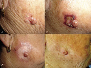
The combination may be more efficacious than imiquimod or cryosurgery alone for the treatment of Bowen’s disease. In a double-blind, placebo-controlled study 16 weeks of daily imiquimod achieved clinical clearance in 9/12 cases of Bowen’s disease after 50 weeks follow-up (13). Moreover, an earlier study with the same imiquimod regime reported histological clearance in 15/16 Bowen’s disease lesions with no relapses in 13/16 patients after 6 months (14). In a smaller series, 8–12 weeks of daily imiquimod accomplished sustained remission of up to 3 years in 3 patients with 4 Bowen’s disease lesions (15). Concerning the efficacy of monomodal cryosurgery, an earlier retrospective study reported a recurrence rate of only 1/128 Bowen’s disease lesions after two freeze-thaw cycles with 30 s freezing time each (3), yet subsequent randomized studies generally report significantly lower clearance rates and concomitantly higher relapse rates of 10–36% with this modality (7, 8). In the present study although, 5 of our patients had therapeutically challenging facial Bowen’s disease lesions (2), the outcome of the treatment was excellent (Fig. 1). This can be attributed to adequate compliance, which was probably facilitated by: (i) shortened total imiquimod application and (ii) milder cryosurgery (2 freeze-thaw cycles, 10–15 s freezing time each in 10/11 lesions) compared with corresponding monotherapies.
Fig. 1. Immunocryosurgery for Bowen’s disease in patient 1 (Table I). (A) Bowen’s disease on the right mandible. (B) The lesion after 3 weeks treatment with imiquimod, just before the cryosurgery session. (C) The lesion 3 months after the end of treatment. Hypopigmentation without scarring is evident. (D) At 24 months follow-up the treated area is re-pigmented, with a reasonable aesthetic outcome. Note the selective anti-neoplastic nature of immunocryosurgery: the biopsy proven dermal naevus that was initially almost encircled by the Bowen’s disease and was included in the treatment field was not appreciably affected by the treatment.
ACKNOWLEDGEMENTS
This study was partially supported by Special Research Account Program, University of Ioannina, number 22195.
The authors declare no conflict of interest.
References