Eleni Sotiriou, Antonia Manousari, Zoi Apalla, Ioannis Papagarifallou and Demetris Ioannides
First Dermatology Department, Aristotle University Thessaloniki, GR-54645 Thessaloniki, Greece. E-mail: elenasotiriou@yahoo.gr
Accepted August 20, 2010.


Eleni Sotiriou, Antonia Manousari, Zoi Apalla, Ioannis Papagarifallou and Demetris Ioannides
First Dermatology Department, Aristotle University Thessaloniki, GR-54645 Thessaloniki, Greece. E-mail: elenasotiriou@yahoo.gr
Accepted August 20, 2010.
Leukaemia is considered the most common malignancy in childhood. However, congenital leukaemia (CL) is extremely rare, accounting for only 0.5–1% of all recorded cases of childhood leukaemia (1–3). CL is defined as leukaemia that is present at birth or develops within the first month of life (1, 4, 5). Leukaemia cutis (LC), which is characterised by infiltration of the skin by immature malignant haematopoietic cells, is a common feature in CL, occurring in up to 25–30% of cases (1, 4, 6). CL and congenital LC (CLC) have similar sex distributions, with a 2:1 male-to-female ratio (1, 3).
Aleukaemic LC is a term applied to describe a form of leukaemia in which infiltration of the skin by leukaemic cells occurs before the appearance of the disease in the peripheral blood or bone marrow (6). Aleukaemic CLC is rarely reported in the literature.
Case report
A 15-day-old boy, born by vaginal delivery at 40 weeks gestation after an uneventful pregnancy, was referred to us for evaluation of a widespread eruption over his trunk and extremities. The exanthema consisted of numerous firm red-to-violaceous subcutaneous papules and nodules, primarily affecting the posterior and anterior parts of the trunk, as well as the proximal regions of legs. The lesions ranged from 1 to 4 cm in diameter. Symmetrically distributed moist scaly erythematous plaques were also observed over the inguinal creases, mimicking napkin dermatitis (Fig. 1). The results of a systematic physical examination were otherwise unremarkable. According to the patient’s medical history, most of the lesions were present at birth and had gradually deteriorated since. Differential diagnosis included nodular mastocytosis, histiocytosis and LC.
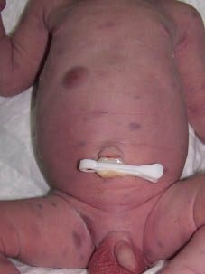
Fig. 1. Firm violaceous subcutaneous nodules on the trunk and legs.
The results of laboratory investigations, including haematology, chemistry, an immunoglobulin assay, a Coombs test, serology for hepatitis A, B, C and TORCH (toxoplasma, other infections, Rubella, cytomegalovirus, HSV), and urinalysis, were normal. Ultrasonography and X-rays also revealed nothing unusual. Bone marrow in two subsequent aspiration biopsy samples did not demonstrate leukaemic infiltration.
Histological examination of a suspicious skin nodule demonstrated diffuse infiltration of the dermis by atypical medium-sized cells with round or folded nuclei and indistinct nucleoli. Immunohistochemical analysis revealed diffuse staining of neoplastic cells for MPO, lysozyme, CD68 and CD43. Staining for CD34, c-kit/CD117, mast cell tryptase, S-100, CD1α, LCA, CD20/L26, CD79α, CD45RO/UCHL1, CD3, CD4, CD8, CD56, CD30/Ki-1 and CD15/LeuM1 was negative. Collectively, these observations provide evidence of cutaneous infiltration by immature myeloid tumour cell blasts. Therefore skin involvement as the primary clinical manifestation of acute myeloid leukaemia (AML) was diagnosed.
The patient’s disease progressed rapidly and he died within 7 days of diagnosis. The ultimate cause of death was established during an autopsy as AML, based on a bone marrow blast population of greater than 20%.
Discussion
Factors associated with CL include inherited genetic traits, environmental factors and factors related to gestation.
Diagnostic criteria for CL include: presentation at birth or within the first four weeks of life; proliferation of immature myeloid, lymphoid or erythroid cells; infiltration of these cells into extra-haematopoietic tissues; and absence of diseases that provoke leukaemoid reactions mimicking CL (1, 4, 5).
The disease presents clinically with hepatosplenomegaly, anaemia, leukocytosis and central nervous system and skin involvement. CLC may precede other manifestations of leukaemia by as much as four months (1). In general, cutaneous manifestations of leukaemia are classified into two groups: non-specific lesions, containing non-leukaemic cells (leukaemids), and specific lesions with true leukaemic infiltrations (LC). Leukaemids may result from immunologic responses to tumour antigens and appear as haemorrhagic lesions, generalised pruritus, exfoliative erythroderma, pyoderma gangrenosum, urticaria, erythema multiforme, erythema nodosum, panniculitis, hyperpigmentation or morbilliform eruptions. LC has a wide variety of unusual presentations and it has been reported to mimic several skin disorders (2, 7). It most commonly presents as multiple, firm, freely movable violaceous, blue or red nodules approximately 1 to 2.5 cm in diameter, giving a “blueberry muffin baby” appearance. There have also been reports of tumours, papules, macules, plaques and ulcers (6). The colour of the lesions is also highly variable. The number of lesions ranges from one (1, 4) or a few to very many. Widespread involvement sometimes affects up to 70% of the body. Oral and ocular involvement is quite rare (1).
The pathogenesis of CLC remains unclear. One possible explanation is that it reflects the early onset of intrauterine leukaemia, since embryonic haematopoiesis begins in the undifferentiated mesenchyme. Alternatively, LC may represent a metastatic infiltration of the skin subsequent to undetectable primary bone marrow disease (1, 8).
Differential diagnosis includes dermal erythropoietic disorders (e.g. congenital infection or haemolytic disease of the newborn), neoplastic infiltrative disorders (especially histiocytosis), and leukaemoid proliferation (transient myeloproliferative disorder) (1, 2, 9).
Biopsy of a suspicious skin lesion is required in order to confirm the diagnosis of LC. Routine haematoxylin-eosin staining reveals a dense, infiltrate, comprising a linear array of pleomorphic leukaemic cells, in the reticular dermis. Despite these suggestive histological characteristics, subtyping of the leukaemic infiltrate almost always necessitates staining for immunophenotypic markers and their correlation with cytologic and cytogenetic data.
While cutaneous manifestations are associated with a very poor prognosis in adults, they do not appear to have any prognostic impact in CL (1). Despite great progress in the treatment of childhood leukaemia, overall survival at 24 months in CL is only 20 to 23% (1–4, 10). Left untreated, it is usually fatal, with almost all infants dying within two months (1). A few isolated cases have undergone temporary or permanent spontaneous remission, even without chemotherapeutic intervention (1, 4, 9). Together with the fact that aggressive chemotherapy and bone marrow transplantation can result in high rates of morbidity and mortality, such cases render the decision whether to treat or not somewhat controversial. Some authors recommend that, in the absence of disease progression or an 11q23 translocation, CL can be managed conservatively with the expectation of spontaneous remission (2).
References

