A Polycyclic Annular Erythematous Plaque on the Trunk: A Quiz
Julie Lemeray, Bernard Guillot and Olivier Dereure*
Department of Dermatology, University of Montpellier 1, Hôpital Saint-Eloi, FR-34000 Montpellier, France.* E-mail: o-dereure@chu-montpellier.fr
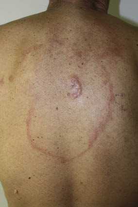
A 57-year-old man was referred for evaluation of an infiltrated erythematous, polycyclic ring of approximately 1 cm in width that had gradually extended in a centrifugal way (Fig. 1) over the preceding months. Physical examination revealed an asymptomatic firm infiltrated 2 cm-wide purple plaque at the centre of the annular element; this lesion had first appeared 4 years earlier and had slowly enlarged unnoticed since then, with no medical advice requested by the patient. He had no systemic symptoms at presentation and clinical examination revealed no palpable lymph nodes or organomegaly. The annular lesion was initially diagnosed as a fungal infection and treated with a fungicidal cream, but no improvement was noted. The central plaque was then submitted to histological examination that showed a dense infiltration of dermis consisting of small-sized lymphocytes with a follicular pattern. Immunostaining showed that this infiltrate was CD20+, focally CD10+, bcl6+, bcl2–. Three subsequent biopsies, of which 2 were performed on the peripheral ring, displayed the same pattern.
Fig. 1. Purple polycyclic ring of subacute evolution surrounding the initial nodular lesion.
What is your diagnosis? See next page for answer.
doi: 10.2340/00015555-1241
A Polycyclic Annular Erythematous Plaque on the Trunk: Comment
Acta Derm Venereol 2011; 91: XX–XX.
Diagnosis: Annular primary cutaneous follicle centre lymphoma
Primary cutaneous follicle centre lymphoma (PCFCL), a low-grade malignancy, is the most common subtype of primary cutaneous B-cell lymphoma. It is characterized by an indolent clinical course and a favourable outcome with a 5-year survival rate of more than 90% (1). Although cutaneous recurrences are common, systemic spread is very rare (1). Clinically, it usually presents as erythematous or purple plaque(s) or nodule(s), which are either solitary or limited in numbers and principally localized on well-defined regions, such as the face, the scalp or the trunk (1). Multi-regional and diffuse forms are less frequent. In rare cases, the clinical presentation may be misleading, as it can mimic another cutaneous disease, a situation that can result in a significant delay in diagnosis. Histologically, the tumour can display a follicular, follicular and diffuse, or purely diffuse infiltrate of predominantly follicular centre small or large centrocytes. The neoplastic cells constantly express CD20, bcl-6 and inconstantly CD10 or CD21, but multiple myeloma 1 (MUM1) is never expressed. The t(14;18) translocation and bcl-2 expression are often absent and the presence of one or both of these markers must raise the possibility of nodal follicular lymphoma with secondary skin localization (2).
According to World Health Organization (WHO) recommendations (2), initial evaluation includes a comprehensive medical history and clinical review of systems, a complete blood cell count, lactate dehydrogenase (LDH) dosage, serum electrophoresis, and a Borrelia burgdorferi serology (along with PCR on skin biopsy in endemic areas) since an association with Borrelia infection has been reported in some lesions (3). GeneScan analysis in search of a dominant clone in skin and in peripheral blood and a computed tomography (CT) scan of chest, abdomen, pelvis (and neck if skin lesions on head or neck area) must be performed as well to ensure the primary cutaneous nature of the lymphoma. Positron emission tomography (PET) remains optional in current recommendations.
For isolated, small-sized, lesions, therapy options include local radiotherapy or excision, although intra-lesional interferon-alpha or intra-lesional rituximab can also be discussed. In case of large and/or multifocal PCFCL, treatment mainly consists of local or regional radiotherapy or intravenous rituximab alone. In some cases, Rituximab-Cyclophosphamide/Vincristine/Prednisone (R-CVP) or Rituximab-Cyclophosphamide/Adriamycin/Vincristine/Prednisone (R-CHOP) chemotherapy can also be used, especially in disseminated or relapsing diseases (2).
Because of the large size of the annular lesion, radiotherapy was not considered in our patient and he was treated with 4 courses of rituximab (375 mg/m2/course), resulting in complete clinical remission of the skin lesions. No recurrence was noted at a 6-month follow-up.
References