


Toshiyuki Aoki
Skin and Allergy Clinic for Itchy Diseases, 2nd Floor, Tennoji Crystal Building, 8–15, Daido-1, Tennoji-ku, Osaka, Japan. E-mail: toshiyuki.aoki@nifty.ne.jp
Accepted Apr 25, 2013; Epub ahead of print Nov 6, 2013
In addition to annular lesions, which are seen in various diseases (1, 2), a disease of unknown cause, called annular erythema in infancy, has been reported independently in several cases (3–8). I describe here 2 typical cases of annular lesions in infantile atopic dermatitis (AD), a condition which I have observed in several cases, but for which there are no published reports.
CASE REPORTS
Case 1
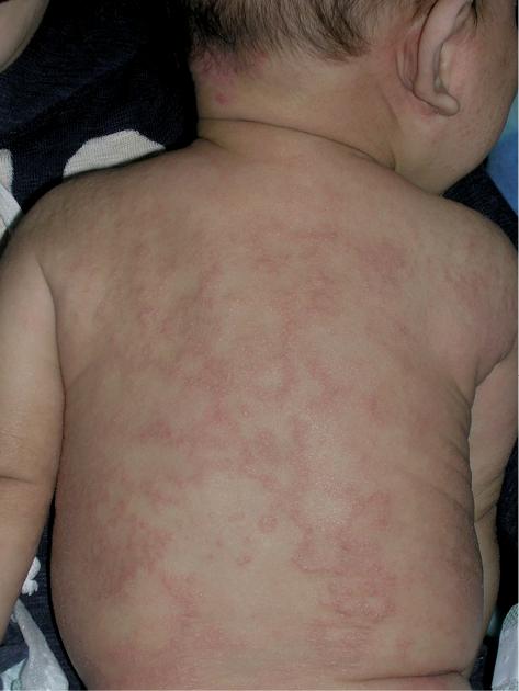
A 2-month-old breastfed boy with no family history of atopic disease first visited us in February 2008. He had dry erythematous papules on his head, forehead, cheeks, and extremities. Large nummular lesions with a bright red border were noted around the shoulder and elbows. Blood analysis revealed values that were within normal ranges, with the exception of hypereosinophilia (1,643/mm3; normal: 50–400/mm3 for all ages), elevated immunoglobulin E (IgE) (147 IU/ml; normal: < 1 year: < 20 IU/ml, 1–2 years: 20–30 IU/ml) and positive radioallergosorbent test (RAST) and positive prick tests to milk (and later to egg white, too). The patient was otherwise healthy. A diagnosis of infantile AD was made, and the administration of a weak topical steroid was started, with the restriction of milk and later eggs in the mother who continued breastfeeding till 8 months. The mother used the steroid only on scratched lesions. The eruptions on the face soon ameliorated, but dry erythematous plaques gradually spread over the trunk and limbs. In April, a group of multiple annular or polycyclic lesions developed on the patient’s back in a mesh-like appearance (Fig. 1), on which there was no sign of scratching. The annular borders moved outward with time, forming larger rings or losing their ring formation, and in May they formed a waste-thread-like pattern, then resolved without trace in June.
When the patient was examined again at just less than 2 years of age (in November), only small dry papules with mild scratching were noted on the trunk and outer limbs, and there were no annular lesions. At follow-up at 4 years of age (in February), a mild goose-flesh-like appearance and dry skin with minimal scratching were observed on the trunk and limbs. The patient had a normal eosinophil count, normal total IgE and negative RAST at this time.
Fig. 1. Case 1, 4 months of age (in April). Mesh-like annular lesions on the back 2 months after the first visit.
Case 2
A 13-month-old boy, the third child of parents with no family history of atopic diseases, presented in March 2008 with a history of eczema that had started soon after birth and had been treated by a paediatrician with strong and mild steroids. He had dry erythematous lesions on the cheeks, chin, jaws and outer aspects of the lower legs. Small and large nummular erythematous and papular lesions were noted on his shoulder, chest, flank and outer surfaces of the forearms. In addition, he had many large erythematous annular macules with raised borders and central clearing on his back, some of which had begun to overlap, resulting in polycyclic forms (Fig. 2). No sign of scratching was noted on these lesions.
There was a slight elevation in total IgE (47 IU/ml) for his age, and hypereosinophilia (673/mm3) was detected. He was RAST-positive to milk, egg white, egg yolk and soy, as well as prick-positive to milk, boiled egg white and boiled soybean. Restriction of cow’s milk, chicken eggs and soy foods was continued.
His family initiated intensive treatment with an antibiotic-containing weak steroid in combination with an antihistamine, and all of the eruptions, including the annular lesions, cleared before June. Towards the end of the year, however, dry skin on the cheeks, and small red papules on the shoulders and chest, were observed. By the following January annular lesions were again observed on the back and the side of the chest. Round-to-annular lesions appeared for the third time 2 years later (in February).
Fig. 2. Case 2, 13 months of age (in March). Typical annular lesions on the back at the first visit.

DISCUSSION
These 2 patients showed typical rashes of infantile AD, and had hypereosinophilia and food allergy. Annular and nummular lesions were noted somewhere on the body at the first visit. In Case 1 a peculiar mesh-like pattern composed of many annular lesions developed later, and in Case 2 multiple annular lesions were noted at the same time with typical atopic lesions, both on the back. These lesions are partly similar to previously reported “annular erythema of infancy”; however, there is no description of AD in the previous reports to exclude a Netherton syndrome case (8). The lesions we observed were composed not only of erythema, but also of papules, and therefore it seems pertinent to call them “annular erythema and papules”.
It is common knowledge that AD is an itchy disease; however, the important feature of “annular erythema and papules” is its low level of itchiness. There were no scratch marks on the annular lesions, and the infants showed no signs of scratching these lesions.
Nummular eczema is known to be associated with xerotic skin (9). Coexistence with nummular eczema may suggest that annular erythema and papules could also be a manifestation of xerotic skin. Recent discovery of loss of function mutation in filaggrine gene (10) has shown that AD may be related to xerotic skin via reduction of natural moisturizing factors.
Whether “annular erythema and papules” is part of infantile AD or a rare association remains unclear. However, this subject provides an interesting topic for future investigation.
ACKNOWLEDGEMENTS
The author would like to thank Ms K. Otsu for her excellent secretarial assistance and Nurse N. Fujimoto for taking photographs of the patients. I also thank Dr K. Ito who referred case 1 to us.
REFERENCES

