


Nicolas Kluger, Sari Koskenmies, Leila Jeskanen, Meri Övermark and Olli Saksela
Department of Dermatology, Skin and Allergy Hospital, Helsinki University Central Hospital, Meilahdentie 2, PO Box 160, FIN-00029 Helsinki, Finland. E-mail: nicolaskluger@yahoo.fr
Accepted May 29, 2013; Epub ahead of print Oct 3, 2013
Permanent tattooing, has gained tremendous popularity for the past 20 years among the Western population. In Europe, the prevalence of tattoos is estimated around 10% (1, 2), with the highest prevalence among the 20–35 years old (25%) (2). In Finland, it is currently estimated that 15% of the 20–30-year-old individuals are tattooed (3). Tattoo inks are currently a combination of organic dyes, metallic salts and various additives including solvents, such as isopropanol, and preservatives (4). Several publications have shed light on possible toxic or carcinogenic compounds that could be introduced in the skin or develop in situ as by-products under various conditions (such as UV light or laser exposure) (5–8). It is only recently that European countries and the council of Europe have started to take actions regarding the tattoo ink marketing, especially by withdrawing some inks that contained potential hazardous components from the market (9). However, the composition of tattoo inks is still not subjected to a strict homogenous regulation. Also, there is currently no test available to assess the safety of inks for the purpose of tattooing. Despite accumulation of the dye to local lymph nodes, which is a well-known consequence of tattooing (7, 10), the potential local and systemic carcinogenic effects of tattoos and tattoo inks remain to date unclear. We report here two additional cases of melanoma that developed on tattoos in two Finnish patients.
CASE REPORTS
Patient 1. In June 2006, a 61-year-old Caucasian Finnish man presented with an inflammatory ulcerated tumoural plaque of the right thigh overlying an old tattoo. Upon examination, a superficial, extensive, heterogenous and asymmetric pigmented lesion underlied and surrounded the tumour lesion (Fig. 1A). According to the patient, the pigmented lesion evolved during the past 5 years. Complete physical examination and full body computed tomography scan were normal. Excision of the lesion confirmed the diagnosis of superficial spreading melanoma with a nodular component (Breslow thickness 15 mm, Clark level IV). Sentinel lymph node exploration was negative. No relapse had occurred before 2009, after which no information was available.
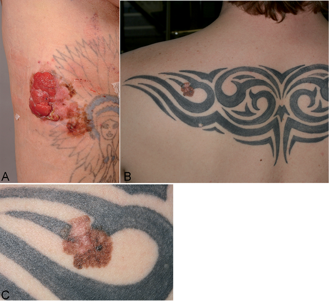
Fig. 1. Case 1. Inflammatory ulcerated tumoural plaque of the right thigh overlying an old tattoo: nodular melanoma overlying a superficial spreading melanoma (A). Case 2. Superficial spreading melanoma within a tribal tattoo of the back (B). Close up view of the lesion (C).
Patient 2. In May 2012, a 32-year-old Caucasian Finnish man presented with a 1.3 cm brown, polychromatic, asymmetric lesion on the upper back within a large black tattoo performed a couple of years earlier (Fig. 1B and C). The patient acknowledged that a small naevus pre-existed before tattooing and gradually changed during the following years. However, careful examination revealed that only the borders of the pigmented lesion reached the tattoo drawing. Physical examination was otherwise normal. Pathology of the surgically removed mole confirmed the diagnosis of a non-ulcerated superficially spreading type melanoma (Breslow thickness 0.4 mm, Clark level II). Tattoo pigments were mainly located on the upper part of the papillary dermis, mainly around capillaries, on both spared edge of the excision margins. Very few dark pigments were found at the very same location as the tumoural area, confirming that the tattooist had most likely avoided tattooing over the initial pigmented lesion. The patient has been symptom-free for 12 months.
DISCUSSION
From 1938 until now, approximately 50 cases of skin cancers have been reported on tattoos, including 16 melanomas (for review see ref 11). Currently, the development of melanoma and non-melanoma skin cancers on tattoos is still considered as fortuitous. This position is supported by several arguments: (i) the rather low number of reported cases compared to the number of tattooed individuals worldwide; (ii) as the prevalence of tattoos and melanoma both increase in the general population, among the young especially, the risk of a coincidental lesion increases as well. In other words, the risk of having a coincidental melanoma on a tattoo increases with the surface of skin that is tattooed; (iii) the absence – so far – of cases of multiple melanoma occurring within the same tattoo, which would be a strong signal for a link, in the absence of any other risk factor for melanoma. Another indirect argument against a link can be taken for our cases. Indeed, if we consider that tattoo inks do contain carcinogenic compounds, one would expect a quantitative effect, i.e. the more ink that is tattooed in the skin, the higher the concentration of carcinogenic components, and the higher the risk of cancer. However, the tattoo of the first patient was only made of thin drawings with no shadings or filling (Fig. 1A), therefore the concentration of tattooed pigments was here quite low. In our second case, it was impossible to determine whether the preexisting mole was already a de novo melanoma or a naevus that transformed secondarily. Some tattoo pigments were found within the melanoma on the sides of the lesion, most likely coincidentally, because the malignant process has extended slowly towards the tattoo.
Although, the incidence of skin cancer on tattoos remains low (11), a notable exception concerns keratoacanthomas (12, 13) known for its proneness to develop quickly within a recent traumatised area (14). It should also be noted the curiosity that keratoacanthomas have mainly been reported within the red parts of tattoos (11), while most of the cases of melanomas have developed within dark tattoos, as in our cases. Indeed, Regensburger et al. (8) found aromatic polycyclic hydrocarbures in some black inks, some of them being classified as carcinogen 2B. However, we think that, rather than having a true and direct carcinogenic effect, dark tattoos may simply mask the clinical malignant modifications and delay clinical diagnosis. The ugly duckling sign is more difficult to detect and, lastly, in our experience, dermoscopy is more difficult because of the exogenous pigmentation (15).
It is highly debatable to suggest to any person wishing to have a tattoo to go to a dermatologist first. However, patients with numerous moles, atypical mole syndrome and familial history of melanoma should definitely seek advice to a dermatologist before tattooing (15). Similarly, tattooists should be careful before tattooing a patient with numerous moles. They should be educated that, in doubt, they should delay the procedure and refer the customer for advice to a dermatologist.
Incidentally, we strongly recommend that the pathologists always use the ICD code for tattoo pigmentation (2013 ICD-10-CM diagnosis code L81.8) in case of any tumour arising on a tattoo so that future epidemiological studies regarding the risk of cancer on tattoos can be performed easily in national cancer registries.
The authors declare no conflicts of interest.
REFERENCES

