


1Department of Dermatology and Allergology, Hannover Medical School, Hannover, 2Department of Dermatology, University of Cologne, Köln, 3Department of Dermatology and Allergology, Medical University of Lübeck, Lübeck, and 4Dermatology and Allergology, Department of Human Medicine, Carl von Ossietzky University of Oldenburg, Oldenburg, Germany
Bullous pemphigoid (BP) is characterized by substantial skin and blood eosinophilia as well as intensive pruritus. Since the pruritogenic cytokine interleukin (IL)-31 is increased in inflammatory skin diseases the aim of this study was to determine whether IL-31 plays a role in BP. Using immunofluorescence, IL-31 expression was analysed in eosinophils derived from blister fluids and skin from patients with BP and IL-31 levels in blister fluids, serum and culture supernatants were determined by enzyme-linked immunoassay (ELISA). High levels of IL-31 expression were observed in BP blister fluids, but they were only marginally elevated in BP serum compared with healthy controls. Eosinophils from either BP blister fluids or skin biopsies showed strong expression of IL-31. Furthermore, peripheral blood eosinophils from patients with BP, but not healthy controls, released high levels of IL-31, reflecting those in blister fluids. In conclusion, eosinophils are a major source of IL-31 in BP and this cytokine may contribute to itch in patients with BP.
Key words: bullous pemphigoid; IL-31; itch; pruritus; eosinophils.
Accepted Apr 24, 2018; Epub ahead of print Apr 24, 2018
Acta Derm Venereol 2018; XX: XX–XX.
Corr: Ulrike Raap, Dermatology and Allergology, Department of Human Medicine Klinikum Oldenburg AöR, Rahel-Straus-Straße 10, DE-26133 Oldenburg, Germany. E-mail: ulrike.raap@klinikum-oldenburg.de
Bullous pemphigoid is an autoimmune disease of the skin and is associated with severe itch and blister formation. In this study we show that eosinophils are a major source of IL-31, a factor that is known to cause itch in other skin diseases, and that IL-31 levels were highly expressed in blister fluids from patients with bullous pemphigoid. We also revealed that eosinophils from patients with bullous pemphigoid released higher levels of IL-31 than eosinophils from healthy
donors. These observations indicate that targeting IL-31 may have a therapeutic potential in bullous pemphigoid.
Bullous pemphigoid (BP) is one of the most common autoimmune blistering skin diseases, with a high prevalence in the elderly population. BP is characterized by circulating IgG and IgE autoantibodies against the hemidesmosomal proteins BP180 and BP230. In most patients with BP autoantibodies recognize the non-collagenous 16A domain (NC16A), located at the membrane-proximal region of BP180. It has been suggested that the disease activity in patients with BP can be determined based on the serum level of these autoantibodies (1). ELISAs, highly specific and sensitive for detecting circulating autoantibodies against the BP180 NC16A region, have been established for the diagnosis of BP (2, 3). It is usually distinguished from other pemphigoid diseases by a combination of direct and indirect immunofluorescence, which show in situ deposits of IgG and C3 at the epidermal basement membrane, together with the clinical appearance of blisters (4). Histopathological examination of lesional skin from patients with BP shows that the majority of sub-epidermal blisters are filled with an eosinophil-rich leukocyte infiltrate (5). Elevated levels of various cytokines and chemokines, including tumour necrosis factor alpha (TNFα), interleukin (IL)-6, IL-8, IL-15, and CC chemokine ligand CCL18 have also been measured in sera and blister fluids of patients with BP, which also correlate with disease activity (6–9). Clinically, erythematous patches or urticarial lesions may precede bullae formation by several days to months. Itch is one of the major symptoms; it presents as mild or severe in almost all patients (10, 11).
IL-31 is a cytokine that plays an important role in itch-associated inflammation. In a transgenic mouse model overexpressing IL-31, mice developed severe itch with skin lesions accompanied by inflammatory cell infiltration and increased numbers of mast cells (12). The phenotype described in IL-31 transgenic mice shows similarity with the clinical picture of patients with atopic dermatitis (AD) (12). In patients with AD, IL-31 serum levels are increased and correlate with disease severity (13–15). Meanwhile, several studies have reported increased IL-31 concentrations in various inflammatory skin diseases, such as chronic spontaneous urticaria, allergic contact dermatitis and mastocytosis (14–19).
IL-31 signalling requires the input of a heterodimeric receptor composed of the IL-31 receptor A (IL-31RA) and the oncostatin M receptor (OSMR) (20). These IL-31 receptors are expressed on several immune cells, including T cells, keratinocytes, dendritic cells, eosinophils, basophils, macrophages and dorsal root ganglia (16, 21–25). Functionally, IL-31 leads to the release of proinflammatory cytokines in human monocytes, macrophages and keratinocytes (26), as well as the release of Th2-type cytokines, including IL-4 and IL-31, in human basophils (25).
In a previous study we have shown that peripheral blood eosinophils are able to secrete IL-31, the level of which was higher in eosinophils from patients with AD compared with non-atopic donors (24). This finding suggests a role of eosinophils in the regulation of IL-31 and, subsequently, of itch in inflammatory skin diseases. Since eosinophilia and itch are 2 main characteristic features of BP, the aims of this study were to clarify whether IL-31 is elevated in patients with BP and whether BP eosinophils are a source of this cytokine.
BP was defined by specific clinical features, including strong blisters on the skin, histopathological findings of subepidermal blistering, a positive indirect and direct immunofluorescence and detection of specific auto-antibodies related to BP180 and BP230. Patients with BP (n = 22, mean age ± standard deviation (SD) 82.3 ± 8.9 years, see Table I for subject characteristics) did not receive systemic immunosuppressive treatment, diaminodi-phenylsulfone, or local therapy with glucocorticosteroids during a 4-week period before taking blood, blister fluid and skin samples. For control subjects we included healthy skin from non-atopic individuals with normal total IgE levels and no allergen-specific IgE-antibodies to 10 common aeroallergens (n = 11). Furthermore, we included 16 patients with mastocytosis as positive controls for IL-31 serum levels. Fresh blisters from the skin from patients with BP (n = 14) were punctured and analysed for eosinophil counts (Neubauer haemocytometer; LO – Laboroptik Ltd, Lancing, UK) and preparation of cytospins (1 × 10⁵ cells/spot). Serum and blister fluids were centrifuged and supernatants were stored at –80°C until processing for further analysis. In addition, serum samples from patients with mastocytosis and controls were handled in the same way. For further preparation, embedded skin was cut into 5-µm thick slices and placed on object slides. Peripheral venous blood was obtained from patients with BP and control subjects (Co) in order to isolate eosinophils and process them for cell culture and prepare cytospins (27).
None of the participants received systemic or topic immunosuppressive treatment. The study was approved by the medical ethics committees of the Hannover Medical School and the Department of Dermatology at the Medical University of Cologne, Germany.
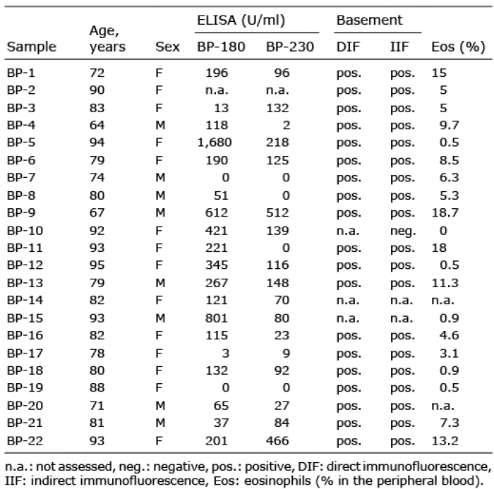
Table I. Subject characteristics of patients with bullous pemphigoid (BP) (n = 22)
Peripheral blood eosinophils were prepared as described previously (24). Briefly, blood was layered on a Ficoll density gradient (Biochrom AG, Berlin, Germany). After removing the interphase and the supernatant, erythrocytes in the pellet were lysed with a lysis buffer (155 mM NH4Cl, 10 mM KHCO3, 0.1 mM EDTA). Residual cells were washed with phosphate-buffered saline (PBS) and eosinophils were isolated by negative immunomagnetic bead selection with CD16 microbeads (Miltenyi Biotech, Bergisch Gladbach, Germany) according to the manufacturer’s protocol. The purity of the eosinophils was > 98%, as assessed by flow cytometry (CD66b/CD16) and Kimura staining. Viability was greater than 99%, as assessed by trypan blue dye exclusion. Isolated peripheral blood eosinophils were used to perform cytospins and cell culture experiments. To perform cytospins eosinophils (1 × 10⁵ cells) were washed in PBS and resuspended in 150 µl PBS. Cell suspensions were centrifuged onto slides (500 rpm, 5 min) using a Cytospin-3 cytocentrifuge (Shandon Southern Instruments, Sewickley, PA, USA). In addition, isolated eosinophils were processed for cell culture. Briefly, eosinophils (2 × 10⁵) were resuspended in culture medium (RPMI 1640 with 10% heat inactivated foetal calf serum (FCS), including 2 mM L-glutamine, 10,000 U/ml penicillin and 10 mg/ml streptomycin, Seromed, Biochrome AG) and cultured for 24 h at 37°C, 5% CO2 in 96-well plates. The supernatants of these unstimulated eosinopihls were used to analyse various cytokine and chemokine levels.
Paraffin-embedded biopsies from lesional skin of patients with BP were analysed by double immunofluorescence using a Vectastain kit (Vector Laboratories Inc., Burlingame, CA, USA), as described previously (17). Before staining, formalin-fixed lesional skin and controls were deparaffinized and treated with antigen unmasking solution (Vector Laboratories). Thawed cytospins from eosinophils of the blister fluids from BP patients were washed with PBS. Cytospins and deparaffinized sections were permeabilized and blocked with 0.03% H2O2 for 5 min at room temperature (RT), followed by the Avidin/Biotin Blocking Kit, according to the manufacturer’s protocol (Vector Laboratories,), and, finally, buffer containing 5% goat serum. After washing, sections were treated with either rabbit anti-human polyclonal IL-31 and rabbit IgG isotype control (Abcam, Cambridge, UK) overnight at 4°C. After washing, biotin-conjugated goat anti-rabbit antibodies (Jackson Immunoresearch, Baltimore, PA, USA) were added for 30 min at RT, followed by Alexa Fluor 488-conjugated Streptavidin (Jackson Immunoresearch) for 10 min at RT. After washing, the 2 blocking steps were repeated and the sections were treated with mouse anti-human monoclonal EG1 abs (Phadia Ab, Uppsala, Sweden) or murine IgG1 isotype control (Abcam) overnight at 4°C. Following a washing step, sections were incubated with biotin-conjugated goat anti-mouse abs (Jackson Immunoresearch) for 30 min at RT. TexasRed-conjugated Streptavidin (Jackson Immunoresearch) was then added for 10 min at RT. Sections were washed and mounted with Vectashield mounting media with DAPI (Vector Laboratories). Analysis was performed with the immunofluorescence microscope Axiolab (Carl Zeiss, Jena, Germany).
IL-31 levels in blister fluids and serum samples were determined using commercially available ELISA kits (Abcam) according to the manufacturer’s instructions. Analysis was performed by the FLUOstar OPTIMA Microplate Reader (BMG Labtech GmbH, Ortenberg, Germany). Detection levels for IL-31 were in the range 7.8–500 pg/ml, as outlined by the manufacturer.
Since BP is accompanied by strong itch and IL-31 is a known pruritogenic cytokine, this study first aimed to determine the concentration of IL-31 in blood and blister fluids from patients with BP, using ELISA, as well as to assess the number of eosinophils within blister fluids. As positive controls sera from patients with mastocytosis were included, in whom we have previously observed elevated levels of IL-31 (17). Surprisingly, the concentration of IL-31 in the blister fluid of patients with BP (108.5 ± 31.8 pg/ml, p < 0.01) was significantly higher than corresponding levels in the peripheral blood. Interestingly, IL-31 levels in BP blister fluids were comparable with the serum levels from patients with mastocytosis (95.6 ± 16.9 pg/ml). IL-31 serum levels of patients with BP (Fig. 1B, 6.4 ± 1.5 pg/ml, p < 0.001) were significantly lower than the serum concentrations from patients with mastocytosis and only slightly higher than those of healthy controls (1.2 ± 0.4 pg/ml, Fig. 1B).
Since increased levels of IL-31 were found in blister fluids from patients with BP, which also contained a large number of eosinophils (Fig. 1A), it was investigated whether eosinophils can produce and release IL-31. For this, peripheral blood eosinophils of patients with BP as well as non-atopic healthy controls were isolated and the supernatants of cultured eosinophils collected after 24 h. These supernatants contained significantly higher levels of IL-31 from eosinophils derived from patients with BP compared with those from healthy controls (Fig. 1C). Furthermore, IL-31 levels in the supernatants of cultured eosinophils of patients with BP were as high as those in blister fluids of patients with BP (Fig. 1C).
Fig. 1. Elevated levels of interleukin (IL)-31 in blister fluids of patients with bullous pemphigoid (BP). (A) Eosinophil numbers present in the BP blister fluids (B) IL-31 concentrations of blister fluid and sera from patients with BP (n = 14), patients with mastocytosis (n = 16) and healthy non-atopic controls (n = 11) were measured by enzyme-linked immunoassay (ELISA). (C) Isolated peripheral blood eosinophils of patients with BP release higher levels of IL-31 (as determined by ELISA) compared with healthy controls. IL-31 data is shown as mean concentrations ± standard error of the mean (SEM). (**p < 0.01; ***p < 0.001).
The blister fluids of patients with BP were characterized by a high number and almost pure population of eosinophils, as assessed by Kimura staining. From these cells cytospins were prepared and stained for IL-31 and the eosinophil-specific marker EG1. As shown in Fig. 2 all BP blister fluid-derived cells were positive for EG1 in addition to IL-31.
Fig. 2. Eosinophils of bullous pemphigoid (BP) blisters express interleukin (IL)-31. Double immunofluorescence staining of eosinophils derived from the blister fluid of a patient with BP (representative of 2 separate donors). Cytospins were stained for EG1 (red, panel B) and IL-31 (green, panel C). Overlays (panel A) display yellow fluorescence for double-positive cells. Nucleus staining was performed with DAPI (blue).
Lesional skin of patients with BP is characterized by high numbers of eosinophils (28). Thus this study aimed to determine whether these cells also express IL-31. Paraffin sections from lesional skin of patients with BP were prepared and stained for EG1 and IL-31. There was a clear infiltration of eosinophils in BP lesional skin, which also expressed IL-31 (Fig. 3). In addition to IL-31-positive eosinophils, however, IL-31-negative eosinophils and EG1-negative IL-31-expressing cells were also observed in lesional skin from these patients with BP (Fig. 3).
Fig. 3. Eosinophils in lesional skin of patients with bullous pemphigoid (BP) express interleukin (IL)-31. Double immunofluorescence staining of eosinophils derived from lesional skin of a representative patient with BP out of 10 analysed. Paraffin sections were stained for EG1 (red, panel B) and IL-31 (green, panel C). Overlays (panel A) display yellow fluorescence for double-positive cells. Nucleus staining was performed with DAPI (blue).
BP is an autoimmune blistering skin disease accompanied by severe itch. Since IL-31 has been described as a highly pruritogenic cytokine, the question of elevated concentrations of IL-31 in patients with BP arises. Our results clearly demonstrate that BP blister fluids are characterized by strongly increased levels of IL-31, which were as high as serum levels of IL-31 in patients with mastocytosis. Furthermore, we provide evidence for the expression of IL-31 in lesional skin of patients with BP, with a distinct expression of IL-31 in skin tissue and blister eosinophils. Finally, we determined eosinophils as a major cellular source of increased IL-31 levels in blister fluids, since isolated peripheral blood eosinophils were able to secrete substantial amounts of IL-31.
Interestingly, IL-31 serum levels of patients with BP in our study were not substantially increased. At present there are conflicting reports regarding relative IL-31 levels in BP sera compared with healthy controls. Kulczycka-Siennicka et al. (29) reported reduced levels of IL-31 in BP sera, whereas a recent study by Salz et al. (30), which was published at the time our current work was first submitted, reported significant increases. Salz et al. (30) also observed high levels of IL-31 in BP blister fluids, as well as an association with granulocytes (30), which we can confirm, although our study demonstrates for the first time that eosinophils are the main source of this cytokine. While we also observed slight elevations in IL-31 from BP sera these concentrations were at least one order of magnitude lower than those seen in BP blister fluids, and in many cases close to the detection limit of the ELISA assay.
Recent evidence has shown that eosinophils contribute directly to blister formation in BP, since dermal-epidermal separation occurred in an ex vivo skin model following incubation with IL-5-activated eosinophils in the presence of BP autoantibodies (31). Previously, neutrophils were thought to play an essential role in BP blister formation (32), which was ascribed in murine models of BP to elastase release from neutrophils in a BP180 IgG-dependent manner (33). It is currently unclear whether eosinophils impact on neutrophil function in BP; however, we have preliminary observations that suggest that IL-31 induces neutrophil chemotaxis and increases intracellular calcium concentrations in human primary neutrophils (unpublished observations). While further studies are necessary, IL-31 release from eosinophils may amplify the inflammatory responses of neutrophils and other granulocytes.
Eosinophils and eosinophil granule proteins have been found in close proximity to peripheral nerves (34, 35). It has been shown that cytokines secreted by eosinophils induce the branching of sensory neurones isolated from murine dorsal root ganglia (35). Recently, we have shown that eosinophils express and release IL-31, which is highest in inflammatory itchy skin disease, including AD (24). In AD, targeting of IL-31 led to a significant improvement regarding skin inflammation and itch, as assessed using a mAB against IL-31 in a phase I/Ib clinical trial (36).Thus, it could be assumed that IL-31 also plays a role in itch and inflammation in BP. However, whether IL-31 also induces the sprouting of nerves in BP, as recently shown by Feld et al. in other settings, is unclear (37). In the blister fluids of our patients with BP we found an almost pure eosinophil accumulation, which suggests that eosinophils are one of the major sources of the increased IL-31 levels in these blister fluids. Since patients with BP experience a strong itch in lesional skin the increased IL-31 levels in blister fluids may explain this localized itch, although future studies are required to verify whether eosinophil accumulation is indeed associated with increased clinical scores for itch. Our present findings, however, suggest that targeting this pruritogenic cytokine may have therapeutic potential in treating BP.
This work was funded by a DFG grant to Ulrike Raap RA 1026/1-2. Judith Engmann was supported through funds of the Hannover Biomedical Research School DFG, GSC 108.


 Click to show fullsize
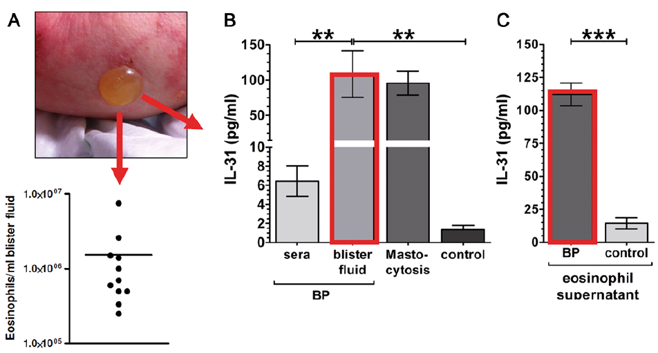 Click to show fullsize
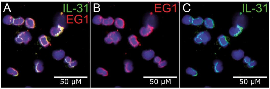 Click to show fullsize
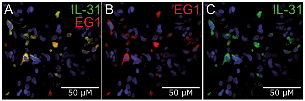 Click to show fullsize