


Dermatology Clinic, University of Catania, Via S. Sofia 78, IT-95123 Catania, Italy. E-mail: cldermct@gmail.com
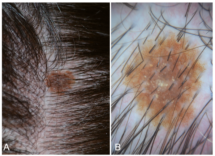
A 7-year-old girl presented with a pigmented lesion on her scalp. The lesion had been present for 2 years and, according to her parents, had changed recently. Clinical examination revealed the presence on the right temporal region of a roundish, smooth, brown macule approximately 0.9×0.7 cm with unusual tiny speckles (Fig. 1A). Dermatoscopy showed a diffuse brown pigmentation and a regular network at the periphery; moreover, several yellowish globules of various shapes and dimensions were observed in the centre of the lesion (Fig. 1B). Reflectance confocal microscopy (VivaScope® 3000, Lucid, MAVIG, Munich, Germany) revealed a meshwork pattern at the dermal–epidermal junction and several roundish clusters of moderately refractive large cells with a hypo-refractive centre in the superficial dermis (Fig. 2A). The lesion was excised and histopathological examination (haematoxylin and eosin staining) revealed the features of a compound melanocytic naevus characterized by junctional nests of large cells with a pale and clear cytoplasm (Fig. 2B).
What is your diagnosis? See next page for answer.
Fig. 1. (A) Roundish, smooth, brown macule with unusual tiny speckles on the scalp of a 7-year-old girl. (B) Dermatoscopy (×10) showing a diffuse brown pigmentation, a regular network at the periphery, and several yellowish globules of various shapes and dimensions in the centre.
Fig. 2. (A) Reflectance confocal microscopy revealing a meshwork pattern at the dermal–epidermal junction and several roundish clusters of moderately refractive large cells with a hyporefractive centre in the superficial dermis. (B) Histopathological examination revealing junctional nests of large cells with a pale and clear cytoplasm (haematoxylin and eosin; H&E).
Acta Derm Venereol 2018; XX: XX–XX.
Diagnosis: Balloon cell naevus
Balloon cell naevus (BCN) is a type of melanocytic naevus that often occurs in young patients, frequently in the head and the neck area. It shows the typical histopathological characteristics of an intraepidermal or compound naevus, along with junctional and/or dermal nests of balloon cells, i.e. large cells with a clear cytoplasm. These cells are formed by the progressive vacuolization of melanocytes resulting from the enlargement and disintegration of melanosomes (1–3). Depending on the number of balloon cells, BCN may either be clinically not distinguishable by other melanocytic naevi or may appear as a yellowish macule or papule generally misdiagnosed as a sebaceous naevus.
The dermatoscopic clue to BCN is the presence of variable number of yellow or white globules (1, 2, 4, 5) that correspond to balloon cell clusters with different grades of melanosome degeneration, along with a regular naevus melanocytic pattern. The presence of yellow globules can also be observed in sebaceous hyperplasia and in some tumours with sebaceous differentiation, such as naevus sebaceous and sebaceous adenoma (1, 6).
Histologically, the main differential diagnosis of BCN is represented by balloon cell melanoma (2, 4, 7), which is an uncommon type of melanoma mostly seen in the head and neck region of middle-aged male subjects. In a review of 34 patients, 2 cases were 19-year-olds and no information on the date of onset was provided, so the existence of balloon cell melanoma in the paediatric age group cannot be excluded (8). Dermatoscopy of balloon cell melanoma has been described in 2 adult cases only, revealing a central structureless white area with a rim of structureless brown and pigmented reticular lines and linear/dotted vessels in 1 case (9), and structureless yellow areas, ulceration and curved/dotted vessels in the other (10); interestingly, no white or yellow globules were observed in either case.
Reflectance confocal microscopy, a new non-invasive imaging technique that displays high resolution, horizontal, en face tissue sections of the epidermis and upper dermis, shows in BCN multiple roundish structures of varying size composed by large cells with a moderately refractive cytoplasm due to the absence of melanin and a hyporefractive nucleus (1, 2, 5).
In conclusion, the presence at dermatoscopy of white-yellowish globules associated with a regular melanocytic pattern should suggest the diagnosis of BCN. Since the presence of these globules alone is not suggestive of malignancy, surgical removal is recommended only when clinical, dermatoscopic or reflectance confocal microscopy signs of atypia are clearly present.


 Click to show fullsize
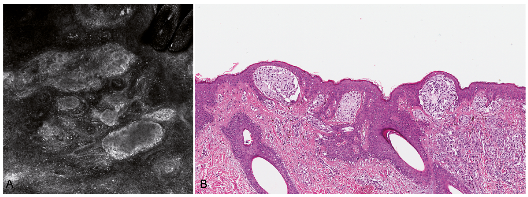 Click to show fullsize