


Department of Dermatology and Allergy Centre, Odense University Hospital, Kløvervænget 15, DK-5000 Odense C, Denmark. E-mail: Kristine.pallesen@rsyd.dk
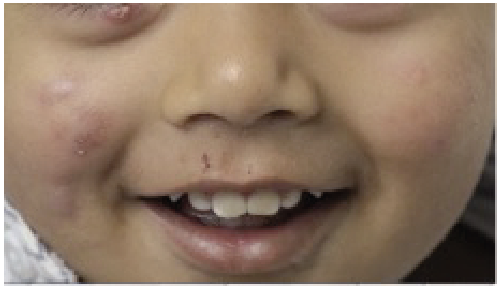
A 1-year-old boy of Afghani origin was referred to the Department of Dermatology due to inflamed nodular lesions on his cheeks. He also had a tendency to bilateral hordeolum on the upper and lower eyelids, which was being followed up by an ophthalmologist.
Three violaceous, small, nodular lesions were observed on the right cheek and similar minor lesions on the left cheek (Fig. 1). He had no lymph node swelling on his neck. Bacterial investigations were negative for atypical mycobacteria.
A skin punch-biopsy from his cheek was performed, and standard histology revealed a stratum corneum with slight hyperkeratosis. Epidermis was hyperplastic with minimal spongiosis. A granulomatous inflammation was accentuated in the reticular dermis.
Treatment with oral penicillin V and topical corticosteroids combined with disinfectants had no effect.
What is your diagnosis? See next page for answer.
Fig. 1. Inflamed nodular lesions on cheeks in a 1-year-old boy.
Acta Derm Venereol 2019; XX: XX–XX.
Diagnosis: Idiopathic facial aseptic granulomas
Idiopathic facial aseptic granulomas (IFAG) are “cold abscesses of the face”. This is a benign, childhood-specific and painless disease characterized by one or more papules or nodules located on the face. The lesions can vary in size (1, 2). The aetiology is unknown, but is thought to be an infantile variant of granulomatous rosacea.
The condition can mimic infantile acne, insect bites, skin infection, sarcoid, granuloma faciale, cutaneous leishmaniasis, dermoid cyst, pilomatrixoma, and other skin tumours.
The dermoscopic features of IFAG were described in a recent report (3). Also, ultrasound has been used to characterize and diagnose this rare condition. It has been reported that both topical and oral metronidazole and topical ivermectin can improve the symptoms of IFAG (2, 4, 5).
The prognosis is good; IFAG will heal spontaneously regardless of the treatment.


 Click to show fullsize