


1Melanoma Unit, Department of Dermatology Hospital Clínic de Barcelona, IDIBAPS, 2Centro de Investigación Biomédica en Red de Enfermedades Raras (CIBERER), Instituto de Salud Carlos III, 3Department of Dermatology, and 4Department of Pathology, IDIBAPS, Hospital Clínic de Barcelona, Barcelona University, Barcelona, Spain
Patients treated with haematopoietic stem cell transplantation are at increased risk of cutaneous malignant neoplasms. There are no reports on the characteristics of melanocytic lesions in patients with chronic graft versus host disease and the value of recognizing these difficult lesions in high-risk patients. The objective of this study is to describe the clinical and dermo-scopic characteristics of melanocytic lesions in patients with chronic graft versus host disease in order to understand their morphology. A prospective cross-sectional study was performed; 10 melanocytic lesions on the trunk and extremities were selected from each patient. A statistically significant association was found between regression and high total dermoscopic score and 7-point checklist score. Lesions were excised or included in short-term digital follow-up. Melanocytic lesions in patients with chronic graft versus host disease developing after allogeneic-haematopoietic stem cell transplantation exhibit marked structural and colour changes similar to melanoma. This is believed to result from the inflammatory process associated with graft versus host disease.
Key words: chronic graft versus host disease; naevi; dermoscopy; digital follow-up; melanoma
Accepted Apr 2, 2019; E-published Apr 2, 2019
Acta Derm Venereol 2019; XX: XX–XX
Corr: Susana Puig, Melanoma Unit, Dermatology Department, Hospital Clinic Barcelona, IDIBAPS, Villarroel 170, ES-08036 Barcelona, Spain. E-mail: susipuig@gmail.com, spuig@clinic.cat
Patients undergoing hematopoietic stem cell transplantation have an increased risk of late complications including skin cancer. The diagnosis of melanoma and atypical melanocytic lesions in patients with chronic cutaneous graft versus host disease is challenging, as the alteration of the skin associated with the disease may mimic malignant dermoscopic findings in benign melanocytic naevi. We evaluated 110 melanocytic lesions and found a significant association between signs of regression and a high score of malignancy. We believe that digital follow-up and comparative analyses reduce the number of excised melanocytic lesions which exhibit marked structural and colour changes similar to melanoma.
Patients undergoing haematopoietic stem cell transplantation (HSCT) are at increased risk of late complications. Survival of these patients has improved over time, but is associated with the development of secondary malignacies, including cutaneous malignant neoplasms. In particular, non-melanoma skin cancer is clearly increased in this group of patients, as is melanoma, and together, these may represent up to one-third of all malignancies in these patients (4).
Chronic graft versus host disease (cGVHD) is a high-risk complication of allogeneic HSCT. In cGVHD there is an activation of T-cell lymphocytes, with consequent inflammation of the skin and other organs producing different clinical manifestations. The muco-cutaneous manifestations of cGVHD can be divided into sclerodermiform and non-sclerodermiform (5). The cutaneous inflammation induces changes in melanocytic lesions and architectural modifications (6). These alterations are responsible for the atypical features in melanocytic lesions of patients with cGVHD and can make clinical diagnosis challenging. In patients with multiple naevi, the comparative analyses approach is used to reduce the number of excisions and detect melanomas, reducing the number needed to treat (7). Dermoscopy improves the diagnostic accuracy of skin cancer and is used as an adjunct to clinical examination in clinical practice. Digital follow-up of patients with atypical mole syndrome reduces the number of skin biopsies of benign lesions and detects early melanoma in these patients (8). However, the impact of dermoscopy and digital dermoscopy has not been described in patients with cGVHD.
Eleven consecutive patients with a personal history of active cGVHD, aged between 18 and 70 years, with more than 50 naevi, who were attending the Dermatology Clinic for GVHD were consecutively included in a prospective cross-sectional study of melanocytic skin lesions. Inclusion criteria were the presence of multiple melanocytic lesions on the trunk and limbs, a previous HSCT, and the presence of active cutaneous cGVHD. Inclusion criteria for the lesions were the 10 largest lesions in the patient, all being larger than 2 mm. Ulcerated or eroded melanocytic lesions and lesions located on special sites (head and neck area, acral sites, and genitalia) were excluded from the study.
Evaluation was performed clinically and via dermoscopy (Dermlite; 3GEN LLC; Salvador Bay, Dana Point, CA, USA), Molemax HD (Derma Medical Systems, Vienna, Austria). Clinical and dermoscopic images were taken using a digital camera (Canon PowerShot G16 (Canon Hongkong Co., Ltd) and a cross-polarized light dermatoscope (DermlitePhoto; 3GEN, LLC).
Evaluation included age, sex, anatomical location, skin type, skin cancer history and number of naevi. Anatomically, the lesions were classified as on the chest, abdomen, back, upper limbs or lower limbs. Dermoscopic features were described according to the third consensus of the International Dermoscopy Society (IDS) (9), ABCD-total dermoscopic score (TDS) (10) and the 7-point-checklist score were assessed (11). For the TDS score (A*1.3 + B*0.8 + C*0.5 + D*0.5) the criteria evaluated were A for asymmetry in 1 or 2 axes; B for 1?8 borders abrupt cut-off; C for colours (white, red, light-brown, dark-brown, blue-grey, black) and D for different structural components (structureless areas, pigment network, branched streaks, dots, and aggregate globules). Lesions with less than 4.75 points were considered benign, between 4.75 and 5.45 points were considered suspicious, and more than 5.45 points malignant. For the classic scoring system of the 7-point checklist, 3 major criteria have a score of 2 points each (atypical network, blue-white veil and atypical vascular pattern) and 4 minor criteria have a score of 1 point each (irregular dots/globules, irregular streaks, irregular blotches and regression structures); lesions with a total score of 3 or more are judged to merit excision. For statistical analysis, the dermoscopic global pattern was divided into 5 groups, as follows: 1: predominantly reticular: reticular, reticular homogeneous, pseudo reticular; 2: predominantly globular: globular, reticular globular, globular homogeneous; 3: homogeneous and unspecific; 4: starburst; and 5: multicomponent. Patients were followed-up every 6 months with digital dermoscopy. Short-term digital dermoscopy follow-up of 3 months in equivocal lesions was performed according to the investigator’s criteria (12).
The lesions were evaluated by a single investigator with experience in dermoscopy and digital follow-up (A.B.).
Excision criteria were: lesions with clear criteria of malignancy; significant changes in short-term digital follow-up; or when the investigators considered the need for excision after comparative analysis of the lesions of every patient (7, 13–15).
Statistical analysis was performed using PASW Statistics 22.0 software (SPSS, Chicago, IL, USA). The χ2 test was used for the comparison of qualitative variables (Fisher’s exact test was applied if any expected cell value in the 2×2 table was < 5). Differences were considered to be statistically significant when p-value was less than 0.05.
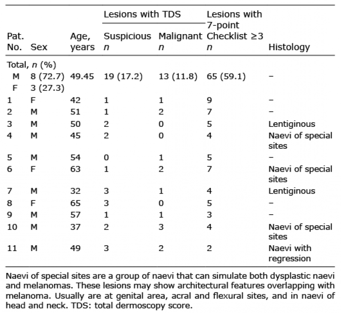
The cohort consisted of 110 melanocytic lesions in 11 patients. The mean ± standard deviation (SD) age of the patients was 49.45 ± 9.6 years at time of inclusion, 72.7% were men (n = 8) (Table I).
Table I. Summary of the lesions included in the study
Three patients had acute myeloid leukaemia, 2 had mantle cell lymphoma, 2 had Philadelphia-positive chronic myeloid leukaemia, 2 had acute lymphoblastic leukaemia, one had follicular lymphoma, and one had multiple myeloma. Five patients received conditioning based on total body irradiation plus cyclophosphamide, while the remaining 6 patients received combination chemotherapy. All patients had received allo-HSCT and only 3 patients were not donor related.
Of the 11 patients evaluated, 6 had both subtypes of cGVHD (lichenoid and sclerodermiform), 3 were lichenoid and 2 were sclerodermiform.
Lesions were located as follows: 50 (45.5%) on the back, 20 (18.2%) on the abdomen, 17 (15.5%) on upper extremities, 13 (11.8%) on the chest, 10 (9.1%) on lower extremities. No lesions from the head and neck area, acral skin, genital mucosa or other special locations as flexural sites were included.
Nine patients had Fitzpatrick skin phototype III and the remaining 2 patients had skin phototype II. Four of the 11 patients had a personal history of skin cancer (2 had basal cell carcinoma and 2 had squamous cell carcinoma). All the patients included in the study had a naevi count of between 50 and 100.
The largest size of the lesions was 12 mm and the smallest 2 mm (mean 3 mm). The codified global dermoscopic pattern was predominantly reticular (reticular, reticulo-homogeneous or pseudoreticular pattern) in 50% (n = 55); homogeneous or unspecific in 36.4% (n = 40); and predominantly globular (globular, globular reticular or globular homogeneous) in 13.6% (n = 15). No lesions with starburst and multicomponent patterns were found.
Dermoscopic evaluation showed dots and globules in 30% (n = 33) of the lesions and 91% (n = 30) of them were atypical. Streaks were present in only 7.3% (n = 8) of the lesions and atypical pigment network in 50.9% (n = 56). Blotching with irregular distribution was present in 20% (n = 22). Regression criteria (blue/grey and white) were seen in 78.2% of the lesions (n = 86) (Table II).
Table II. Frequency of the dermoscopic criteria of the melanocytic lesions evaluated
According to the TDS rule, 19 pigmented lesions with a score of suspicious (17.2%) and 13 (11.8%) with a score higher than 5.45 (classified as malignant) were found. The mean ± SD TDS of the lesions was 3.95 ± 1.4. The 7-point checklist resulted in 59.1% (n = 65) of the lesions with a score of 3 or more and the mean ± SD score was 2.58 ± 1.4.
Blue-grey regression (Fig. 1) was present in all the lesions with a TDS of malignancy (p = 0.024) and in 93.8% of the lesions with 3 or more points in the 7-point check-list (p = 0.000) (Table III). White regression was described in 68.4% (n = 13) of the tumours with “suspicious TDS” (p = 0.000) and in 89.5% (n = 17) of the lesions with more than 2 points on the 7-point check-list (p = 0.002).
Fig. 1. (a) Clinical image of an atypical lesion. (b) Dermoscopy (x30) shows atypical pigment network, structureless areas (arrow) and the presence of regression (blue/grey) at the centre (circled). The total dermoscopic score (TDS).
Table III. Correlation between dermoscopy criteria, total dermoscopy score (TDS) and 7-point checklist
Almost 60% (n = 10) of the lesions with mixed regression (blue-grey regression and white regression) had suspicious TDS (p = 0.007) and 82.4% (n = 14) of lesions with this kind of dermoscopy pattern presented more than 3 points in the 7-point check-list (p = 0.029).
Regarding dots and globules, 93.9% of the naevi with this dermoscopy sign had a high score (>2) in the 7-point checklist (p = 0.000). All the lesions with atypical dots and globules had more than 2 points in the 7-point check-list (p = 0.000).
As regards the score suggesting malignancy in the 7-point checklist, 80.4% of the lesions had atypical pigment network (p = 0.000), 100% of the lesions had atypical streaks (p = 0.000), and 86.4% of the lesions had atypical blotches (p = 0.003).
Rosettes and negative pigment network were not seen in any of the lesions.
Six atypical lesions in 6 patients (5.4% of all the lesions in this study) had a TDS consistent with melanoma and were excised (Fig. 2) with a pathological result of benign naevi with some atypia, inflammation or regression, but none proved to be malignant melanoma. The remaining lesions were included in a short-term dermoscopy digital follow-up of 3 months and were not excised, as no significant changes were observed. Patients were followed-up every 6 months with total body dermoscopy and digital dermoscopy of all included non-excised lesions. None of the patients developed melanoma (mean follow-up 12 months) (Fig. 3).
Fig. 2. (a) Clinical image of the abdomen of the patient. The evolution of this lesion was unknown and it was very different from the others. (b) Dermoscopy (x30) shows an atypical highly pigmented lesion. (c) Large nests of melanocytic cells were seen in the superficial dermis. Fibroplasia and abundant melanophages are also observed (haematoxylin and eosin; HE×100).
Fig. 3. Pigmented lesion with structureless areas (arrow) and regression signs (circled). Short-term digital follow-up showed no changes (30x). The time between figure (a) (b) is 3 months and 6 months between the others.
Patients undergoing HSCT often develop cutaneous cGVHD and will need dermatological care in specialized units for better management and control. These patients have an increased risk for skin cancer (2, 16). Several risk factors for squamous cell carcinoma, basal cell carcinoma and melanoma in HSCT recipients have been identified. The association between cutaneous malignant neoplasm development and cGVHD is poorly understood; however, cutaneous malignant neoplasm often develop at sites of previous cGVHD (17).
There is a lack of systematic description in the scientific literature of melanocytic lesions in patients who develop cGVHD after HSCT. In 2002, Andreani et al. (18) described the number of naevi in allogeneic bone marrow transplantation recipients. They found that conditioning with high-dose chemotherapy, absence of severe cutaneous cGVHD, and young age at transplantation were the main variables that independently predicted an excess of naevi; but they did not describe the dermoscopy of these lesions (18). In another paper, Kaminska-Winciorek et al. (19) reported the dermoscopic features and follow-up of 13 patients with acute GVHD.
The current study describes, for the first time, the dermoscopic characteristics of melanocytic lesions in patients with cGVHD. In our patients we found a high percentage of benign lesions with malignant or suspicious scores in TDS and the 7-point check-list in the same patient. The higher scores of malignancy of lesions in patients with cGVHD are probably due to the inflammatory process of the skin (20). These lesions exhibited significant regression (80%) and atypical pigment network (51%), which are criteria associated with melanoma. Differences were found between both methods (TDS and 7-point checklist), which can be explained by the fact that the 7-point and the TDS score evaluate structures and colours in a different way. Also, in the 7-point checklist the impact of atypical pigment network and regression structures is higher in the final score than in the ABCD rule (10, 11).
Dermoscopy, digital dermoscopy follow-up and comparative analyses are important tools for evaluating difficult melanocytic skin lesions (7, 8). In complicated cases, such as patients with atypical mole syndrome, the comparative analysis is highly sensitive for detecting melanoma. Moreover, digital dermoscopy follow-up has been introduced for the monitoring of patients at high risk of melanoma, such as in atypical mole syndrome, familial melanoma syndrome or patients with xeroderma pigmentosum. Using these methods early melanoma can be detected, thus reducing the number of unnecessary excisions of equivocal benign lesions (8).
The efficiency of digital dermoscopy for the detection of early melanomas in high-risk patients, such as in patients with xeroderma pigmentosum (21, 22), those receiving anti-BRAF treatment (23–25), and atypical mole syndrome, has been assessed. Salerni et al. (8) in a meta-analysis of digital follow-up concluded the utility of short-term monitoring in patients with atypical mole syndrome. Perier-Muzet et al. (24) published a prospective follow-up, by digital dermoscopy, of melanocytic lesions in patients under vemurafenib treatment. They reported that more than 50% of the captured lesions showed changes induced by the drug and that these changes occurred very rapidly (approximately 32.6% by the third month after initiation of treatment). The dermoscopic criteria that led to surgical removal in these cases were: localized changes in focal pigmentation (“dermoscopic islands”), the appearance of dark structureless areas, and changes in network or in diameter. In contrast to our study of cGVHD patients, they concluded that standard recommendations regarding digital follow-up could not be applied in patients receiving BRAF inhibitors.
In the current study the use of comparative analyses and digital follow-up were useful to manage the atypical lesions in patients with cGVHD without excision of many benign naevi. Short-term follow-up did not show significant dermoscopic changes at the 3-month period.
Regarding the histopathology of these lesions, features of naevi were found at special sites in 3 of the 6 biopsies, with the presence of dermal fibroplasia and large nests in the superficial dermis and at the dermal epidermal junction. Extensive regression was found in one lesion. The remaining 2 lesions had a lentiginous pattern of growth. Naevi of special sites or naevi with site-related atypia can show unusual histopathological features that mimic dysplastic naevi and malignant melanoma. However, changes of pseudomelanoma, which are frequently seen in recurrent naevi, or subepidermal clefts of epidermolysis bullosa-associated naevi were not found. The cause of these variations is not known, but it has been suggested that external factors (i.e. trauma, ultraviolet radiation) or intrinsic factors (i.e. age, hormonal factors) could play a role in their development (26, 27).
In conclusion, the diagnosis of melanoma and atypical melanocytic lesions in patients with cutaneous cGVHD patients is challenging as the alteration of the skin associated with the disease induces atypical dermoscopic findings in benign melanocytic naevi. Digital follow-up and comparative analyses (ugly duckling sign) were useful in reducing the number of excisions in our patients. In the future, other modern technologies, such as reflectance confocal microscopy (28–30) or electrical impedance spectroscopy (31) may be useful to improve the accuracy of clinical diagnosis in these difficult cases.
Limitations of this study were the low number of patients, the short follow-up period and the lack of malignant lesions.
The authors thank the patients and their families, who are the main reason for these studies, the nurses from the Melanoma Unit of Hospital Clínic of Barcelona for helping to collect patient data; and Helena Kruyer for helping with English editing and correction of the manuscript.
Funding sources: This research was partly funded by the Instituto de Sanidad Carlos III Support Grant CM17/00042.
The authors have no conflicts of interest to declare.


 Click to show fullsize
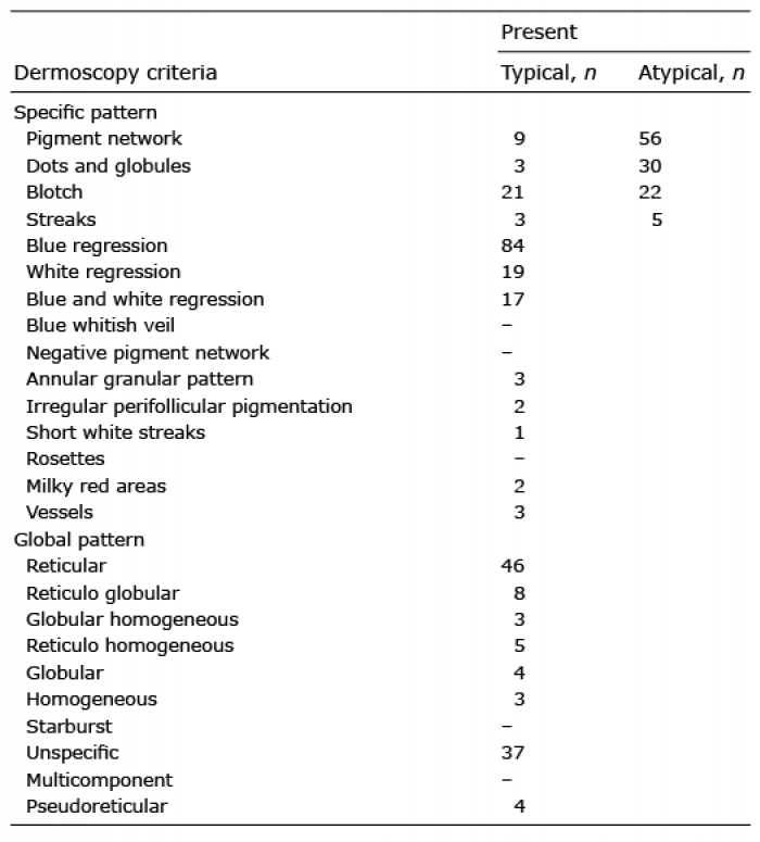 Click to show fullsize
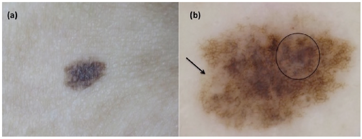 Click to show fullsize
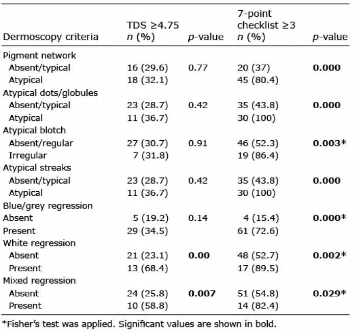 Click to show fullsize
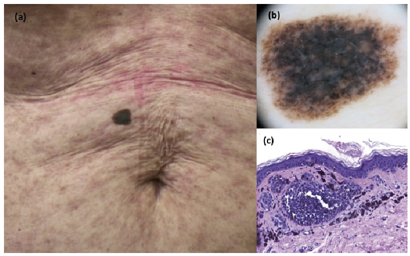 Click to show fullsize
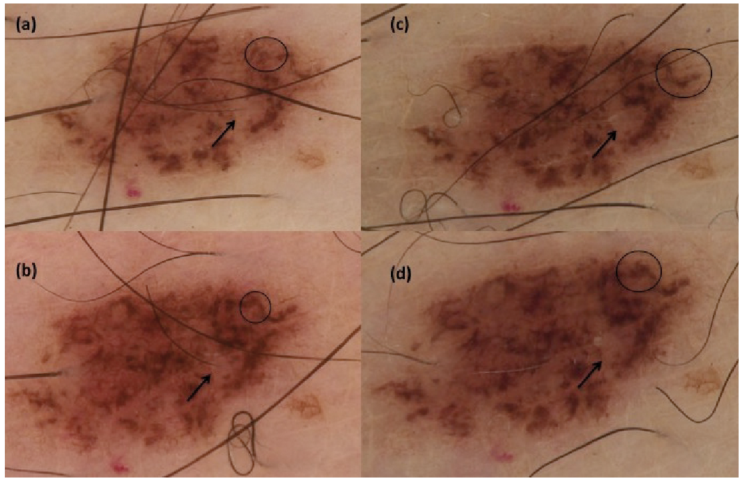 Click to show fullsize