


Department of Dermatology and Allergy Centre, Odense University Hospital, DK-5000 Odense, Denmark. E-mail: rikke.maria.nielsen2@rsyd.dk
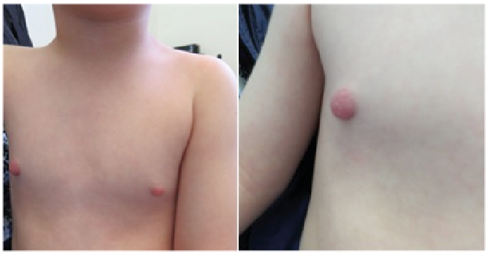
An otherwise healthy 4-year-old boy was admitted to the Department of Dermatology with swelling of his right areola (Fig. 1). The symptoms began 4–5 months earlier with a red spot at the nipple and a rash on the chest. There was no history of tick bite or erythema migrans (EM), fever or malaise. A paediatrician had referred him to ultrasound, which revealed a slightly thickened right mammary papilla with no signs of abscess, haematoma or tumour.
The rash disappeared after use of moisturizing cream, but the nipple became increasingly swollen and red. Therapy with topical corticosteroid was ineffective.
The right areola was enlarged, swollen and indurated when palpated. There were no signs of eczema, secretion, scaling or ulceration. The child did not report any tenderness or itching and appeared to be in good general health.
What is your diagnosis? See next page for answer.
Fig. 1. 4-year old boy with swollen nipple and areola.
Acta Derm Venereol 2019; XX: XX–XX.
Diagnosis: Borrelia lymphocytoma
The clinical appearance was compatible with Borrelia lym-phocytoma (BL), which was confirmed by positive Borrelia burgdorferi IgG antibody (+1). At a check-up when the diagnosis was confirmed, the swelling of the nipple had already decreased. Treatment was initiated with an oral suspension of penicillin 50 mg/ml, 10 ml (500 mg) 3 times daily for 21 days. Further improvement would be expected during the following 3–4 months and no further follow-up was required.
Borreliosis is a vector-borne infection caused by the spirochete Borrelia burgdorferi. The spirochete is transmitted to humans by the bite of an infected tick. Borreliosis is a common tick-transmitted disease in the northern hemisphere, especially in USA and Europe (1). Infection with Borrelia can manifest with symptoms from different organs and there is not always a history of EM, which is the most common clinical presentation of borreliosis (2). Other more rare manifestations are arthritis, carditis, neuroborreliosis or skin manifestations such as acrodermatitis chronica atrophicans or BL (3, 4).
BL is a B-cell pseudolymphoma, which can be seen weeks to months after transmission of Borrelia burgdorferi. It is a rare condition; hence diagnosing can be challenging because of the lack of awareness. Also, the incubation period for BL is usually longer than for EM, which might explain why recall of tick bite is more often missing in children with BL compared with children with EM (5).
In our case, BL was the only clinical presentation of Borrelia infection, as the former rash was not diagnosed as EM. The appearance of BL is usually a painless, bluish-red, soft nodule or plaque (5). The diagnosis is based on patient’s history and clinical findings, but serological tests are recommended to confirm the diagnosis, especially in atypical cases (2).
BL seems to occur more frequently in children than adults (6). Berglund et al. (3) found BL in 7% of children with borreliosis compared with only 2% of infected adults. Interestingly, the location of the lesion can vary between children or adults, as the predilection site for BL seems to be breasts, scrotum, or sometimes ear lobes, in adults (7), whereas it is more frequently located on the ear lobes in children (5, 6). There is also a noticeable geographical difference in the prevalence of BL, as BL is only rarely observed in children and adults in North America, but more often in European children (2). This might be explained by distinct strains of ticks causing BL, which are predominant in Europe (8).
The treatment of BL is oral antibiotics for a period of 3 weeks. The choice of antibiotics is usually phenoxymethylpenicillin, amoxicillin, doxycycline or cefuroxime (2). The child in the current case was treated with oral penicillin for 21 days.


 Click to show fullsize