


1Dermatology Department, 3Pathology Department and 4Gastroenterology Department, Hôpital Saint Louis, AP-HP, 1 Avenue Claude Vellefaux, FR-75010 Paris, and 2Paris VII Sorbonne Paris Cité University, Paris, France. *E-mail: jean-david.bouaziz@aphp.fr
Accepted Oct 3, 2019; E-published Oct 3, 2019
Paradoxical psoriasis (PP) is the most frequent dermatological side-effect of tumour necrosis factor (TNF) inhibitors. Photosensitive psoriasis has rarely been reported to date, and mostly in the paediatric population. We report here an unusual case of sun-induced PP in an adult patient treated with infliximab for Crohn’s disease.
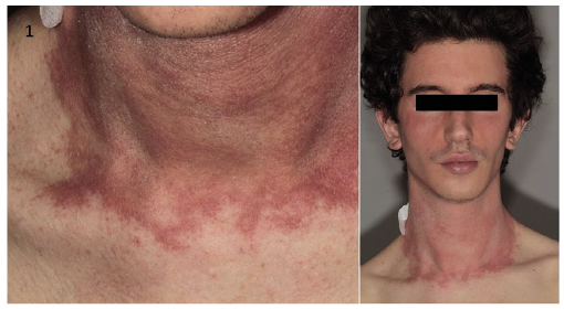
A 23-year-old man, non-smoking, with no significant personal or familial past medical history presented an acute eruption on the face in June 2017. He had been followed up since November 2012 for stricturing Crohn’s disease, reaching the last 30 cm of the ileum and revealed by chronic right iliac fossa pain and constipation. Infliximab (IFX) was started in April 2015 for chronic active disease despite long-lasting steroids use and maintenance therapy with azathioprine. Magnetic resonance enterography after one year of IFX revealed a regression of small bowel thickening and disappearance of parietal enhancement on delayed phase. While the patient was in complete clinical remission, he developed the eruption (Fig. 1). The last infusion of IFX had been administered 3 weeks previously. Dermatological examination revealed an itchy and very well limited erythematous papulosquamous eruption of the face and neck. There was no fever and the rest of the physical examination was unremarkable. The only possible triggering factor was a photo exposition. The patient had not applied plants or topical treatments to the skin. He had not taken any photosensitizing drugs or concomitant therapy for Crohn’s disease.
Fig. 1. Clinical features. Erythematous papulosquamous eruption of the face and neck. Permission given to publish these photos.
Blood tests, including complete blood cells, C-reactive protein, serum electrolytes, creatinine concentration, and liver tests, were normal. On skin biopsy, the epidermis was acanthotic with elongation of the rete ridges and parakeratotic. It was also spongiotic with migration of lymphocytes. The dermal infiltrate was composed of lymphocytes and few neutrophils. These features were suggestive of PP (Fig. 2). Due to the eruption severity, IFX was discontinued and topical tacrolimus was introduced. Skins lesions dramatically improved after 2 weeks and completely healed after 8 weeks. There was no recurrence of psoriasis lesions after a follow-up of 2 years.
Fig. 2. Histopathology of the lesions. Psoriasiform epidermal hyperplasia with perivascular lymphocytic infiltrate in the dermis. Haematoxylin and eosin stain, ×200 magnification.
PP following anti-TNF-α therapy occurred most frequently among women and patients with a personal or familial history of psoriasis, with a predilection for palmoplantar pustulosis and increased incidence of psoriatic arthritis (1–4). In a French single-centre observational cohort of 583 patients, the prevalence rate of PP was 10.1% after a mean duration of 38.5 months of treatment. Anti-TNF-α was discontinued in 18.6% of cases (2). In this study, young age and duration of the involved anti-TNF-α agent were significantly associated with onset of PP. After switching to another anti-TNF-α agent for PP, a recurrence occurred in approximately half of the patients (2).
The pathogenesis of this reaction remains unclear. It has been suggested that interferon-α overproduction by plasmacytoid dendritic cells, increased number of interleukin (IL)-17/IL-22 secreting Th17 cells and IFN-secreting Th1 cells may play a crucial role (4, 5).
Photosensitive psoriasis is a rare entity with only one series of 20 patients reported to date (6). It usually occurs during summer, is characterized by a major female predominance, possible occurrence in paediatric patients, association with a familial history of psoriasis, other photosensitivity symptoms and abnormal response to phototherapy. To date, evidence-based guidelines to manage PP are lacking due to insufficient data. One case of remission of photosensitive psoriasis successfully treated by IFX has been reported previously (7).
Photo-induced autoimmune diseases, such as lupus-like syndrome and dermatomyositis, phyto photodermatitis, substance or drug photoallergic dermatosis, could induce an identical eruption. Our patient had not applied plants or topical treatments to the skin, such as topical retinoids, and had not taken photosensitizing drugs, such as tetracycline antibiotic, sulphonamides, or tricyclic antidepressants. Among the others facial dermatosis, the semiology of the eruption was not in favour of severe rosacea or seborrhoeic dermatosis. The patient had no past history of photosensitivity, which is not compatible with a polymorphous light eruption. Thus, the 2 remaining diagnoses were photo-induced autoimmune diseases and PP. The histological aspect observed in our patient is suggestive of PP.
No patch testing was performed because no attributed molecule was found. However, this may be a limitation of the study.
To our knowledge, this is the first reported case of possible photosensitive PP induced by anti-TNF-α therapy.


 Click to show fullsize
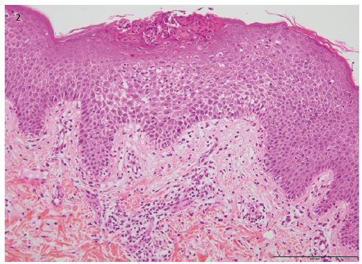 Click to show fullsize