


Department of Dermatology, Medical Faculty and Medical Center – University of Freiburg, Freiburg, Germany
The term skin fragility disorders describes a group of conditions in which the structural integrity of the skin is compromised and its resistance to external shear forces diminished. Skin fragility can have different causes, ranging from genetic variations to inflammatory or physical phenomena. The genetic skin fragility disorders, collectively called epidermolysis bullosa, serve as a paradigm for the study of causes and mechanisms of skin fragility. Recent biomedical research has revealed substantial genetic heterogeneity of the epidermolysis bullosa group, delivered ample new knowledge on its pathophysiology, and facilitated the design of evidence-based therapeutic strategies. The therapy development process extends from in vitro testing to preclinical validation in animal models, and clinical trials. This article reviews different approaches to curative and symptom-relief therapies, and appraises their status and perspectives for clinical implementation.
Key words: skin blistering; genodermatosis; molecular therapy; symptom-relief.
Accepted Dec 18, 2019; Epub ahead of print Feb 6, 2020
Acta Derm Venereol 2020; 100: adv00053.
Corr: Leena Bruckner-Tuderman, Department of Dermatology, Medical Faculty and Medical Center – University of Freiburg, Hauptstrasse 7, DE-79104 Freiburg, Germany. E-mail: bruckner-tuderman@uniklinik-freiburg.de
The term skin fragility describes skin that blisters and breaks easily upon mild friction or trauma. Skin fragility can have many causes, ranging from genetic variants to a compromised immune system, infections or adverse drug reactions. Studies of genetic skin fragility disorders, such as epidermolysis bullosa, have provided better understanding of their causes and mechanisms. At least 20 genes may be involved in epidermolysis bullosa, and secondary phenomena, such as inflammation or fibrosis, can worsen the disease. No cure is yet available, but international research is developing novel approaches to cure the disease and alleviate its symptoms. This article reviews these new developments and appraises their clinical implementation.
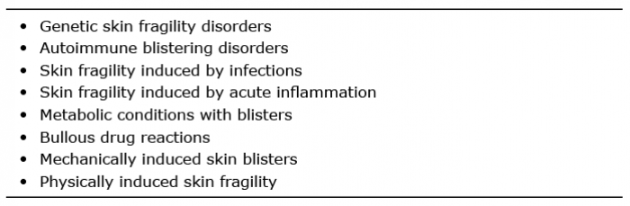
The term skin fragility refers to pathologically altered skin that blisters and breaks easily upon mild friction, pressure or trauma. The breakage can occur in different skin layers, within the epidermis, along the dermal–epidermal junction, or in the upper dermis. The factors that can cause skin fragility and blistering range from genetic variations to (auto)immune, inflammatory, physical, mechanical, infectious, or drug-induced processes. Correspondingly, many classes of disorders can be described using this term, and the differential diagnosis is broad (1) (Table I). As a genetic skin fragility disorder, epidermolysis bullosa (EB) serves as a useful paradigm for these disorders, and research into EB has delivered new information about the pathophysiology of skin fragility that is clinically relevant (2, 3). For example, molecular characterization of autoantigens in acquired blistering diseases has led to the development of molecular diagnostic tests that are in standard use in diagnostics, management and monitoring of autoimmune bullous disorders (4, 5).
Table I. Differential diagnosis of skin fragility
EB has been studied intensively, and the genetic causes and disease mechanisms of the different EB types are rather well understood (1–3). The initial simple assumption that a single pathogenic gene variant/mutation explains all symptoms still holds true in principle. However, the complexity of cellular and molecular processes unleashed by mechanical stress on EB skin is far greater than anticipated; a fact that has major consequences for the design and development of therapies.
As background for the discussion and appraisal of therapy developments, a short introduction to EB, its current diagnostics and management follows.
Epidermolysis bullosa classification
The EB group encompasses 4 main types: EB simplex (EBS), junctional EB (JEB), dystrophic EB (DEB), and Kindler syndrome (6) (Table II). The division into types is based on the morphological level of separation within the dermal–epidermal junction zone. In EBS, the blisters form within the epidermis, in JEB within the basement membrane, and in DEB just below the basement membrane. In Kindler syndrome, blisters can form at all levels. A common hallmark for all EB types is trauma-induced skin blistering and fragility, but each of them contains a number of subtypes, in which the extent of skin lesions and the associated organ manifestations can vary to a great extent (Fig. 1). In April 2019, an international EB consensus classification meeting took place in London. Experts from all over the world updated and revised the consensus classification; the new classification paper is in preparation (6). The main changes are related to the EBS group that has expanded significantly in the past 5 years. Some of the very severe forms in this group, but also mild disorders with minimal skin fragility, were clearly regarded as skin fragility disorders, but not as EB. The new classification includes for the first time syndromal EB subtypes with multi-organ involvement.
Table II. Major types of epidermolysis bullosa (EB)
Fig. 1. Typical clinical presentations in different types of epidermolysis bullosa (EB). (A, B) EB simplex (EBS). (A) Blisters, erosions and scaling in the foot of a 2-year-old child. (B) Disseminated blisters on the trunk and extremities of a newborn. (C, D) Junctional EB (JEB). (C) Blisters, erosions and loss of nails in the hand of a 7-year-old girl with moderate JEB. (D) Typical extensive skin fragility in the buttocks area and back of a newborn with severe generalized JEB. (E, F) Dystrophic EB (DEB). (E) Strong scarring and fusion of digits in the hand of an 8-year-old girl with severe generalized DEB. (F) Trauma-induced blistering, inflammation and scarring on the shins of a 12-year-old girl with moderate DEB.
Modern diagnostics of epidermolysis bullosa
A well-defined diagnosis, with as much molecular precision as possible, is recommended for all patients with EB. A clear diagnosis facilitates disease management, including prognostication and genetic counselling (7, 8). Furthermore, as novel therapies emerge, molecular diagnosis is often a prerequisite for inclusion in clinical trials; it will also be needed for application of future personalized therapies (8). The recommended diagnostic procedure involves immunofluorescence mapping of a skin biopsy as a first step; this enables identification of the blistering level and definition of candidate gene(s) for subsequent genetic analysis. In cases with inconclusive clinical presentation, genetic diagnostics using next generation sequencing (NGS) technologies, such as EB gene panel-based diagnostics or clinical exome analysis, are recommended (7).
Current disease management
Since there is currently no cure for EB, a combination of symptomatic treatment modalities is used, depending on needs. Protection from trauma, cleaning, disinfecting, and moisturizing the skin belong to daily basic measures. Different wound management modalities are defined in guidelines (http://www.debra-international.org/clinical-guidelines). Furthermore, since involvement of other organs is common in more severe EB, and since chronic skin fragility and painful wounds diminish the quality of life of the affected individuals and their families, interdisciplinary and multi-professional management, including psychosocial care, are highly recommended (www.debra-international.org/clinical-guidelines/complete-eb-guidelines.html).
Expert centres and European Reference Networks
Numerous expert centres for EB exist worldwide. Most of these are members of the EB-Clinical Network “EB-Clinet” (www.EB-Clinet.org), which works together with the patient groups (www.debra-international.org). The centres provide information and advice to patients and caregivers, as well as services ranging from diagnostics to genetic counselling and interdisciplinary management plans. In 2017, the European Commission launched European Reference Networks (ERNs) for rare diseases for high-quality diagnostics, management, and research. The goal is to tackle complex or rare diseases with a concentration of knowledge and resources (https://ec.europa.eu/health/ern). The ERNs provide a dedicated IT platform, telemedicine tools and a virtual advisory board of specialists from different disciplines to evaluate the diagnosis of a patient and plan the treatment. An important principle is that the medical knowledge and expertise “travel”, and not the patients, who should have the comfort of staying at home in their supportive environment. ERN-Skin encompasses 56 healthcare providers from 18 countries who are endorsed by their national authorities and committed to pool their knowledge and expertise within the framework of the ERN-Skin (https://ern-skin.eu/). Two approaches are taken: (i) a disease approach with 8 sub-thematic groups on high-level patient management and research; (ii) a transversal approach focusing on teaching and training, E-health, registries and research, deep phenotyping and clinical outcomes. One of the 8 sub-thematic groups deals with EB.
Despite all the structural developments in the field of rare skin diseases, the unmet medical need remains high, and novel evidence-based therapies are urgently needed. Development of new treatments is strongly promoted by patient advocacy groups, which are very active in setting priorities and funding patient-oriented research (www.debra-international.org; www.ebresearch.org/, www.cure-EB.org).
As the therapeutic era for skin fragility disorders progresses it becomes clear that therapy strategies with “intention to cure” are far more complex and difficult than expected. Gene therapy development faces technological challenges with vectors, targeting skin stem cells, achieving long-term therapeutic effects, etc. Therefore, a variety of methodologies relating to gene replacement, gene editing, and modifying transcription and translation are being tested. Because patients demand more rapid development of treatments that bring relief, the focus has turned to so-called symptom-relief and regenerative therapies that, although they do not bring cure, will alleviate symptoms, offer relief and improve quality of life. The therapies that have reached a clinical trial stage and are recruiting trial participants are summarized in Table III.
Table III. Currently recruiting clinical therapy trials for epidermolysis bullosa (EB) (as of June 2019)
Gene therapies
Retrovirus-mediated gene correction in keratinocytes and subsequent grafting of gene-corrected epidermal sheets was developed many years ago as a principally valid method to treat JEB or DEB skin (9, 10 and references therein). Recently, this method was used to replace approximately 80% of the skin surface in a very severely ill child with JEB (9, 10). A similar approach is being tested in DEB for maintenance of wound healing (11). So far, 7 patients with RDEB have been treated with COL7A1-gene corrected keratinocyte grafts, many of them have durable wound-healing (www.abeonatherapeutics. com). However, the classical gene therapy approaches still deal with technological issues relating to vector safety and to optimal transfection/transduction efficiency of stem cells. Gene editing using the CrispR/Cas technology has shown promise in correcting COL7A1 mutations in RDEB keratinocytes (12) and RDEB fibroblasts (13) in vitro and at a preclinical level. Further research strategies encompass approaches with gene-corrected iPS cells (14–17). A newly introduced technology employs a non-integrating, replication-incompetent herpes simplex virus 1 (HSV-1) vector expressing human collagen VII (www.krystalbio.com). The vector preferably targets keratinocytes/epidermis, and a pilot trial using topical treatment of DEB addresses wound-healing as a primary outcome marker (Table III).
Natural gene therapy
The term “natural gene therapy” describes revertant mosaicism, i.e. the spontaneous conversion of a somatic cell with a mutation and pathological phenotype into a cell that has acquired a second, compensating mutation and gained a normal phenotype (18). Revertant mosaicism is relatively common in genetic disorders, and in most classic EB types revertant mosaic skin patches can be found by a well-trained expert. Approximately 5 years ago, the first “natural gene therapy”-based treatment of EB was reported, JEB skin was transplanted with small split-thickness revertant grafts (19). More recently, cultured epidermal autografts generated from clinically revertant skin were applied to treat DEB wounds in 3 patients. The take was 55–87%, and the clinical effects remained for at least 76 weeks of follow-up (20).
RNA-based therapies
Different approaches can be used to skip or replace exons at the RNA level. In an ex vivo RNA trans-splicing-based approach 7 exons were replaced, including the one with a KRT14 mutation, to correct the cellular phenotype in EBS keratinocytes. The corrected keratinocytes formed a stable epidermis in a xenograft model, indicating that trans-splicing-mediated RNA therapy could have potential for clinical implementation (21). Another option is to employ antisense oligonucleotides to skip the mutated exon in the transcription process. Subsequently, a polypeptide that lacks the amino acid sequence encoded by the skipped exon is synthesized; this is usually at least partly functional. Collagenopathies are particularly suitable for this approach, since exons of collagen genes are typically in-frame and small. Their deletion is not likely to cause major structural changes in the affected protein. Of the EB genes, the collagen VII gene is interesting, since exon 73 harbours a high number of mutations. In vitro experiments showed that antisense oligonucleotide-induced skipping of exon 73 leads to a partially functional collagen VII that could potentially improve DEB skin functions (22, 23). A phase 1/2 multicentre clinical trial plans to test this approach in DEB patients carrying specific mutations (www.wings-tx.com).
Premature termination codons read-through
The idea of read-through of premature termination codons (PTC) arose from the knowledge that nonsense-mediated mRNA decay is often caused by PTC (24). Overriding the mutation during transcription would presumably generate a full-length translation product, i.e. a polypeptide with a minor modification that is likely to be adequately functional. Aminoglycoside antibiotics induce PTC read-through. However, the neighbouring nucleotides of the mutations influence the efficiency of the read-through and, therefore, not all PTC are suitable for aminoglycoside treatment. Gentamicins suppressed COL7A1 and LAMB3 mutations with some efficacy in vitro and in vivo (25, 26). Human therapy trials assess the suitability and tolerability of intravenous gentamicin in RDEB and topical gentamicin in JEB (Table III). A challenge with this category of drugs is the spectrum of adverse effects, such as renal and ototoxicity, or potency to induce contact sensitization. Gentamicin B1, a minor gentamicin constituent, has been suggested to be superior in this context due to its high potency to suppress PTC and its low toxicity (27). Amlexanox, an anti-inflammatory drug, can also induce PTC read-through. In vitro, in collagen VII-negative DEB cells with PTC mutations, it induced collagen VII protein production (28).
Protein therapy
Protein therapies, in particular enzyme replacements, have been designed and tested for several inborn errors of metabolism (29). In case of EB, the challenge is that many of the proteins that are mutated and/or missing (collagens, laminins, keratins) are large and, by the nature of their physiological functions, have a tendency to form aggregates. These characteristics do not facilitate intravenous administration and homing of the protein to the required site of action. With this background it seems surprising that intravenous and intradermal injections of recombinant collagen VII in DEB model mice resulted in homing of some collagen into the skin and the dermal–epidermal junction, without major adverse effects (30). A clinical trial is currently testing the safety of recombinant collagen VII in RDEB (Table III; http://phoenixtissuerepair.com).
With increasing experience in preclinical and clinical development of therapies for EB, the complexity of treatment-related issues has surprised most scientists (8, 31). We realize that curative therapies will need many years to enter the clinics and, at the same time, the pressure from patients for treatments increases. The scientific community has reacted by searching for possibilities to modify disease activity and to alleviate symptoms. The rationale for such symptom-relief approaches comes from basic research on disease mechanisms in EB. Many in vitro and preclinical studies have laid the foundation for using cells or targeting, for example, cytokines or growth factors that drive EB phenotypes (8). The goal of these treatments is to improve functions of the skin and make the patients feel better. Three groups of symptom relief therapies are delineated below: (i) regenerative cell-based therapies; (ii) topical pharmacological therapies; and ( iii) systemic therapies with biomolecules and repurposed drugs.
Regenerative cell-based therapies
From many different angles, cell therapies for EB have turned out to be more challenging than initially expected. They are very unlikely to bring cure, and have recently been re-grouped into the category of disease-modifying treatments. Currently, both local and systemic applications are being tested for disease-modifying capacity.
Intradermal cell injections
Early investigations with intradermal injections of fibroblasts or human bone marrow-derived mesenchymal stem cells into RDEB mice demonstrated that the cells produced collagen VII that homed into the dermal–epidermal junction and ameliorated its stability (32–34). However, in humans the tolerability and efficacy of this therapeutic approach were poorer than expected. The injections were very painful and improvement of the skin very limited (35). One study observed a comparable improvement of wound healing in DEB, regardless of whether fibroblasts or vehicle was injected (36). Recently, the approach has been modified with the use of gene-corrected fibroblasts that produce large amounts of collagen VII. Preliminary information indicates that the injections bring some de novo collagen VII into the treated areas, but the full potential of this approach remains to be seen (37; www.fibrocell.com).
Systemic stem cell therapies
Bone marrow transplantation has been tested as treatment for different genetic diseases, including severe DEB (38). Disappointingly, the therapeutic effect and duration were not as positive as hoped for and, as is well known, the complications of bone marrow transplantation can be life-threatening (39). Subsequently, different conditioning regimens have been tested, most recently a regimen that combines reduced-intensity conditioning, post-transplant cyclophosphamide and infusions of immunomodulatory allogeneic mesenchymal stromal cells (40). Treatment of children with RDEB with intravenously administered human allogeneic mesenchymal stem cells made them feel better, but brought no collagen VII into the skin (41). The efficacy of an ABCB5-positive subpopulation of mesenchymal stem cells for symptom-relief in adults with RDEB is assessed in a current trial (www.rheacell.com). In addition, cord-blood derived stem cells have shown some potential as systemic anti-fibrotic treatment in a preclinical setting (42).
Topical pharmacological therapies
Diacerein from rhubarb root extracts has been implicated as possible treatment for EBS skin (43, 44). The rationale involves the capacity of diacerein to dampen the inflammatory response caused by epidermal cell rupture in EBS (43). The cell disruption is a consequence of keratin 5 and 14 mutations that cause intermediate filament aggregation and loss of stabilization by the cytoskeleton. In vitro data demonstrated both anti-inflammatory properties of diacerein and its potential for stabilizing EBS cells, then a pilot clinical trial demonstrated fewer blisters in diacerein cream-treated skin in part of the study population (44).
Wound-healing in EB can be supported by another plant-derived compound with anti-inflammatory properties, namely betulin-based oleogel isolated from birch bark. Betulin was shown to support keratinocyte differentiation (45), enhance re-epithelialization and facilitate wound healing in vitro and in vivo (46, 47). An ongoing placebo-controlled phase 3 study assesses the efficacy of oleogel in patients with EB, regardless of subtype (48).
Systemic disease modifying therapies
Anti-inflammatory approaches. Recent basic research, followed by preclinical and clinical validation, has revealed an unanticipated role for inflammatory cascades in EB. In EBS, keratin mutations and keratinocyte fragility induce expression of specific cytokines and T-cell-mediated inflammatory responses, which manifest with itch as a bothersome symptom (49, 50). A vicious circle is generated by itch, scratching and subsequent skin blistering, which leads to a stronger inflammatory response. Although non-specific anti-inflammatory therapies with NSAIDs are not beneficial, first pilot studies with specific systemic treatments show promise. For example, anti-IL17 interval therapy with apremilast worked well in 3 individuals with of EBS (50).
Antifibrotic therapy approaches. Based on an ample body of scientific literature, severe DEB can be regarded as a systemic disease, since systemic inflammation is prominent and the secondary progressive fibrosis affects many organs (51). Therefore, drugs that inhibit inflammation and fibrosis could potentially relieve symptoms in DEB, such as inflammation-caused itch or formation of strictures and contractures, including fusion of digits.
A repurposed drug, losartan, has shown such benefits in DEB on the preclinical level (52). This drug for treatment of high blood pressure also has anti-fibrotic potential in some disease constellations. The mechanism is based on its ability to inhibit TGFβ signalling via AT-1 receptor antagonism (52). Since inflammation and hyper-active TGFβ signalling contribute to DEB-associated fibrosis in a major manner (8, 53, 54), losartan appeared suitable as treatment. The expectations were met in losartan-treated RDEB model mice, inflammation and TGFβ activity were reduced, progression of fibrosis inhibited and fusion of digits delayed (53). As a logical next step, a clinical trial currently assesses safety and tolerability of losartan in children with moderate-to-severe DEB. The study is also likely to generate preliminary information on the ability of losartan to alleviate symptoms in human DEB (Table III).
Another modulator of TGFβ signalling is the small leucine-rich proteoglycan decorin. Endogenous decorin levels are known to correlate with clinical severity in RDEB (55). In a preclinical study, systemic administration of lentivirally overexpressed human decorin reduced TGFβ levels and fibrotic traits, and enhanced survival of the RDEB mice (56). These observations indicate that extracellular matrix biomolecules modulating TGFβ signalling may have potential for systemic anti-fibrotic therapy for DEB.
In addition to the above small (bio)molecules, a high mobility group box 1 (HMGB1)-derived peptide may improve systemic fibrosis in DEB. HMGB1 has variable functions and has been implicated in both physiological and pathological processes (57). In the context of EB, its relevance lies in its ability to release a specific anti-inflammatory population of mesenchymal stem cell from the bone marrow into the circulation and from there into damaged skin (58). First treatments of RDEB mice with a HMGB1-derived peptide resulted in improvement of skin fibrosis and gastrointestinal strictures (K. Tamai, personal communication).
The multitude of approaches to EB treatments and the rapid developments of research methodologies raise our hopes that first evidence-based therapies for EB will enter clinics in the foreseeable future. To date, biologically valid treatment modalities for most severe EB types have advanced to preclinical and clinical testing, but all strategies still face substantial challenges, including technical issues, safety considerations, or issues related to practical clinical implementation and the duration of the clinical effects. Many of the pilot studies have made us realize that much work is still needed for better understanding of the disease mechanisms and skin stem cell properties. These must be further elucidated, and new therapeutic targets identified. Based on all we know today, the prediction is that future treatments for EB will represent individualized medicine based on the patient’s mutation constellation, phenotypic characteristics and prominent disease mechanisms. They are likely to encompass combinations of different therapeutic principles: curative and symptom-relief therapies. It is easy to imagine therapeutic regimens using alternating gene, cell and drug therapies to win the best clinical outcomes and to reduce adverse effects. Once therapies are available for wide clinical implementation, the next big challenges will have to be tackled, such as cost and worldwide access to therapy.
The author’s research has been supported for many years by grants from the German Research Foundation DFG, the EU E-Rare Programme, and by Debra International. She has no conflict of interest to declare.


 Click to show fullsize
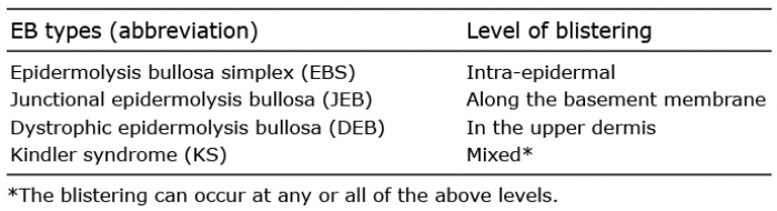 Click to show fullsize
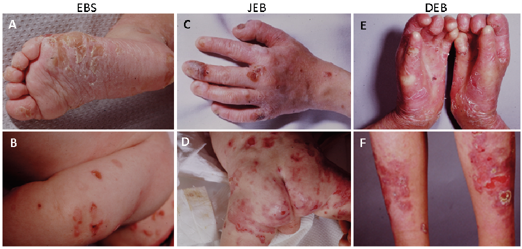 Click to show fullsize
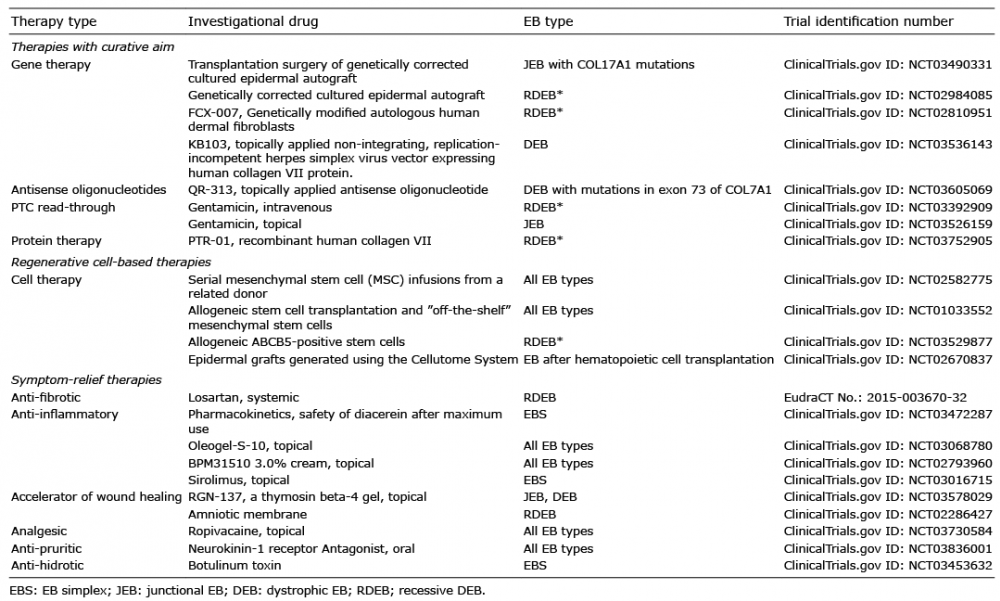 Click to show fullsize