


1Department of Dermatology, Hospital Universitari Sagrat Cor, Grupo Quironsalud, Viladomat Street, 288, ES-08029 Barcelona, 2Department of Dermatology Hospital Universitari Arnau de Vilanova, Lleida and 3Department of Dermatology, Hospital del Mar-Parc de Salut Mar, Barcelona, Spain. E-mail: arcadi.altemir@gmail.com
A 34-year-old woman presented with extensive asymptomatic papules over her upper leg. Physical examination revealed multiple coalescing small brownish papules, 2–5 mm in diameter, with a velvety surface on the anterior and lateral aspect of her left thigh in a grouped segmental distribution (Fig. 1). The patient referred a slow but progressive extension over the last 20 years. She was otherwise healthy and had no family history of similar lesions. A punch biopsy was performed. Histopathological findings revealed an expanded papillary dermis with loosely collagen fibres and scarce cellularity. Alcian blue highlighted an elevated mucin deposit in the papillary dermis. The epidermis showed mild papillomatosis with elongated rete ridges and orthohyperkeratosis (Fig. 2). Expectant management was performed.
What is your diagnosis? See next page for answer.
Fig. 1. Multiple coalescing brownish papules on the lateral aspect of the patient’s left thigh.
Fig. 2. (a) An increase in mucin in the papillary dermis that displace collagen fibres with a mild acanthotic epidermis and elongated rete ridges. Some mature fat cells are also present in the upper dermis (haematoxylin-eosin stain ×100). (b) A prominent increased mucin deposition in the papillary dermis (Alcian blue stain at pH 2.5 ×100).
Acta Derm Venereol 2021; 101: adv00371.
Diagnosis: Mucinous naevus (naevus mucinosis)
Mucinous naevus (MN) is a rare naevoid condition characterized by mucin deposition in the papillary dermis. Since the original description by Redondo-Bellón et al. in 1993 (1), fewer than 30 cases have been reported (2). This condition has classically been considered a rare variant of connective tissue naevus, although some authors have included MN within the spectrum of primary cutaneous mucinoses, or even epidermal naevi (3).
Clinically, MN presents as asymptomatic skin-coloured to brownish papules that slowly grow in confluent plaques following a unilateral or zosteriform distribution. Lesions present at birth or early adulthood and are commonly located in the lower back, predominantly in male patients (2). Histologically, it is characterized by a thickened papillary dermis with an empty appearance secondary to a band-like acid mucopolysaccharides deposition.
Mucin deposits are thought to be secondary to an overproduction of hyaluronic acid (positive with Alcian blue at pH 2.5, and not staining at pH 0.5) by activated fibroblasts (3, 4). Epidermal changes are variable and may be useful to define 2 different histopathological subsets: connective tissue naevi of the proteoglycan (CTNP) type when the epidermis is normal; and epidermal-CTNP type when hyperkeratosis and acanthosis with elongation of the rete ridges is found (5, 6). In addition, a reduction in elastic fibres and collagen can be found in the papillary dermis, as well as a scarce cellular component, which consists of CD34-positive fibroblasts (7, 8).
The unique clinicopathological features of mucinous naevus may help to distinguish this entity from other hamartomatous nevoid lesions and cutaneous mucinoses. The clinical differential diagnosis includes epidermal naevus, connective tissue naevus, and naevus lipomatosus superficialis. However, the presence of mucin deposition in the papillary dermis may permit ruling out these diagnoses. In contrast to mucinous naevus, in cutaneous focal mucinosis, mucin deposits are larger in size and not limited to the papillary dermis (9). Papular mucinosis, also known as lichen myxoedematosus, present glycosaminoglycans deposition, but they are usually deposited at a lower level, tend to occur in older patients, and are manifested by multiple non-confluent lesions, giving rise to a different clinical picture (10).
Due to its benign nature, treatment for mucinous naevus is not required. No spontaneous regression has been reported. Surgical excision or carbon dioxide laser treatment may result in scarring, but may be considered in selected cases only for aesthetic purposes (5).
The authors have no conflicts of interest.


 Click to show fullsize
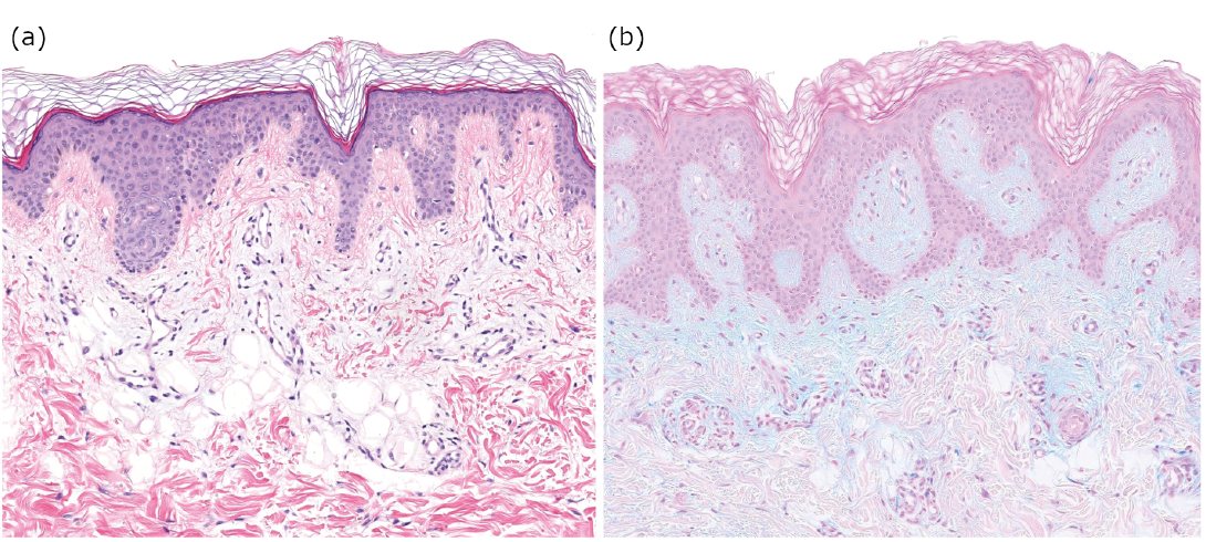 Click to show fullsize