


1Department of Dermatology and Venereology, Institute of Clinical Sciences, Sahlgrenska Academy, University of Gothenburg and 2Region Västra Götaland, Sahlgrenska University Hospital, Department of Dermatology and Venereology, Gothenburg, Sweden
Incomplete excisions of melanocytic lesions occur despite the intention of complete removal. The aim of this study was to determine the incomplete excision rates for benign and malignant melanocytic lesions and the associated risk factors. Demographic, clinical, and histopathological data possibly associated with incomplete excision were collected from 2,782 consecutive excisions between 2014 and 2015. Of these, 269 melanocytic lesions (9.7%) were incompletely excised. Multivariate analysis revealed the following risk factors for significantly higher incomplete excision rates: lesions located in the head and neck area (odds ratio (OR) 3.95, 95% confidence interval (95% CI) 2.35–6.65), surgery performed by general practitioners (OR 3.01, 95% CI 2.16–4.19), the use of a punch excision technique (OR 2.83, 95% CI 1.96–4.08), and excision of non-dysplastic naevi (OR 1.58, 95% CI 1.11–2.23). In conclusion, more caution should be taken when excising melanocytic lesions in the head and neck area, general practitioners require more surgical training, and punch excisions of melanocytic lesions should be avoided.
Key words: incomplete excision; surgery; melanocytic lesion; melanoma; naevus.
Accepted Mar 10, 2021; Epub ahead of print Mar 16, 2021
Acta Derm Venereol 2021; 101: adv00421.
doi: 10.2340/00015555-3784
Corr: John Paoli, Department of Dermatology and Venereology, Sahlgrenska University Hospital, Gröna Stråket 16, SE-413 45 Gothenburg, Sweden. E-mail: john.paoli@vgregion.se
Surgical excision of melanocytic lesions is a common procedure. Incomplete excisions cause unnecessary distress and may lead to further surgery. This study reveals that approximately 1 in 10 melanocytic lesions are incompletely excised. Risk factors associated with higher incomplete excision rates are: naevi and melanomas in the head and neck area; surgery performed by general practitioners; punch excisions, and excisions of non-dysplastic naevi. Thus, larger surgical margins may be required in the head and neck area. Furthermore, general practitioners may need better surgical training in this procedure and updates on surgical guidelines. Lastly, the use of punch excisions for these lesions is not recommended.
Cutaneous melanoma (termed melanoma in this paper) is a potentially lethal skin cancer. Melanoma causes illness and suffering, as well as high societal costs (1). Furthermore, the incidence of melanoma among fair-skinned populations is continuously increasing (2–5). Since surgical excision is the main treatment modality for melanomas (6–9), and distinguishing melanomas from naevi can be challenging, excision of all types of melanocytic lesions is common (10). A recent meta-analysis found that the number of benign lesions needed to be excised to find one melanoma was 22.62 for primary care physicians and 9.6 for dermatologists (11).
According to current guidelines, melanocytic lesions deemed to require removal due to any suspicion of melanoma should ideally be treated initially with primary diagnostic excision (6–9). When possible, the goal of the diagnostic excision should always be complete excision, i.e. no visible tumour cells in the specimen margins upon histopathological examination (6–9). Although a recent meta-analysis has shown that incomplete excisions and partial punch biopsies of melanomas prior to their complete removal do not increase the recurrence risk or decrease survival rates (12), incompletely excised melanocytic lesions with any suspicion of melanoma should be considered a treatment failure. An incomplete excision of a non-dysplastic or low-grade dysplastic naevus does not require a re-excision (13), but if the patient develops a recurrent naevus it may mimic melanoma and lead to diagnostic difficulties in the future, as well as unnecessary re-excisions (14, 15). In the case of high-grade dysplastic naevi, re-excision is recommended, due to the histopathological uncertainty (16). Thus, incompletely excised melanocytic lesions may cause distress for both patients and treating physicians. Previous studies on melanoma report varying incomplete excision rates of approximately 10–24% (17–21). One earlier study, which only included children treated in tertiary care, reported on incomplete excision rates of naevi specifically with 10.8% being incompletely excised (22). However, little is known about the incomplete excision rate of benign and malignant melanocytic lesions as a group.
Several risk factors for incomplete excision of melanocytic lesions have been identified. Firstly, incomplete excision rates for melanoma are higher among general practitioners (GP) than for dermatologists and physicians in other surgical specialties (17, 19, 20). Nevertheless, one report showed similar incomplete excision rates among GPs and other specialties.(21) Secondly, facial location of both melanomas and melanocytic naevi is also a risk factor for incomplete excision (21, 22). In addition, surgical margins, excision type, prior biopsy, and histopathological characteristics can affect the incomplete excision rate for both melanoma and atypical melanocytic lesions (23, 24).
The objective of this study is to determine the incomplete excision rates for melanocytic lesions when complete removal is intended. A further aim was to analyse which risk factors are associated with higher incomplete excision rates.
This retrospective observational study only included data regarding primary diagnostic excisions of melanocytic lesions where the intention was to achieve a complete excision. All lesions were histopathologically verified at the Department of Pathology at Sahlgrenska University Hospital, Gothenburg, Sweden between 1 January 2014 and 31 December 2015. All melanomas (invasive and in situ) and dysplastic naevi excised during the entire study period were included, as well as all non-dysplastic naevi excised between 1 January 2015 and 30 June 2015. Lentigo maligna and lentigo maligna melanomas were excluded, as were melanocytic lesions excised with shave biopsies, since these are known to have an increased risk of incomplete excision (25, 26). Lesions with equivocal histopathological data and cases lacking data on margin control were also excluded. Incomplete excisions were defined as having positive resection margins with visible tumour cells at the specimen border. The project was previously approved by the regional ethics review board in Gothenburg, Sweden.
Data collection
Relevant demographic, clinical, and histopathological data were collected from available patient medical records and histopathological reports. Collected data possibly associated with higher incomplete excision rates were: the medical specialty and experience (specialist or non-specialist) of the physician performing the excision, the clinical surgical margin, the lesion diameter and body site, the histopathological diagnosis and the excision type (i.e. elliptical excision, circular scalpel excision or punch excision).
Statistical analysis
All statistical analyses were carried out with the statistical program “R” version 3.0.3 (The R Foundation for Statistical Computing, Vienna, Austria). Fisher’s exact test was used to compare proportions. Wilcoxon’s rank sum test was used for 2-sampled tests. Kruskal–Wallis test was used to compare 3 or more groups. A multiple logistic regression analysis, adjusting for possible confounders, was performed to identify risk factors that were independently associated with incomplete excision. In order to test for association between 2 categorical variables while testing for a third, the Mantel-Haentzel test was applied. All tests were 2-sided and a p-value of < 0.05 was considered statistically significant.
Histopathology reports of 3,328 lesions from 2,786 patients were reviewed. A total of 546 lesions were excluded. Reasons for exclusions were: other excision types (n = 234), equivocal histopathological data (n = 208), lack of information regarding complete vs incomplete excision (n = 61), and lentiginous melanocytic lesions (43). Thus, a total of 2,782 melanocytic lesions from 2,353 patients were included in the study. The median age of the patients was 51.8 years (range 0.7–97.0 years) and 53.3% were women. The women were significantly younger than the men (median 46.2 vs 57.8 years, p < 0.001).
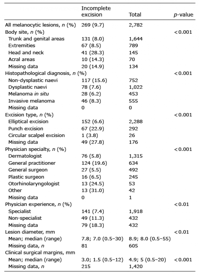
In the univariate analysis, all studied parameters affected the incomplete excision rates significantly. The proportions of melanocytic lesions incompletely excised in relation to risk factors possibly associated with incomplete excision rates are listed in Table I. In total, 269 melanocytic lesions (9.7% of all lesions) including 74 of the 1,008 invasive or in situ melanomas (7.3%) were incompletely excised. Lesions located in the head and neck area were most often incompletely excised compared with other body areas. Non-dysplastic naevi were twice as often incompletely excised compared with the other types of melanocytic lesions. Disregarding otorhinolaryngologists and “other” specialties (paediatric surgeons, ophthalmologists, orthopaedic and maxillofacial surgeons) who performed very few excisions, GPs had the highest incomplete excision rate. Punch excisions were more often incomplete compared with the other surgical techniques. Lastly, incompletely excised lesions as a group had a smaller lesion diameter and were excised with a narrower clinical surgical margin.
Table I. Clinicopathological parameters of the melanocytic lesions and their univariate association with surgical outcome
Ten of the 292 lesions excised with punch excision (3.4%) were malignant including 7 in situ and 3 invasive melanomas (among these, 3 in situ and 1 invasive melanoma were incompletely excised). Approximately half of the punch excisions were performed by dermatologists (51.4%), while the remaining ones were primarily performed by GPs (40.8%). The size of the lesions excised with punch excision was significantly smaller than for elliptical and circular scalpel excisions (median 4 mm for punch excision vs 8 mm and 15 mm for elliptical and circular scalpel excisions, respectively; p < 0.001).
The clinical surgical margins used for non-dysplastic naevi were smaller than those used for dysplastic naevi and melanomas (median 3 mm for non-dysplastic naevi vs 5 mm for dysplastic naevi, in situ and invasive melanomas, respectively; p < 0.001). However, there was a large amount of missing data regarding clinical surgical margins in general, and the amount was larger for non-dysplastic naevi compared with the other melanocytic lesions (76.9% vs 41.5%).
In Table II, clinicopathological characteristics possibly associated with incomplete excision rates are analysed in relation to the 4 specialties performing the majority of the excisions of melanocytic lesions. GPs mostly excised benign melanocytic lesions, but 84 lesions (13.2%) were found to be invasive melanoma or melanoma in situ. GPs used a narrower clinical surgical margin and performed punch excisions more frequently than dermatologists, general surgeons and plastic surgeons.
Table II. Clinicopathological characteristics of the melanocytic lesions grouped by the four most frequent physician specialties carrying out the diagnostic excisions
Based on the findings from the univariate analysis, the clinicopathological variables were regrouped to binary measures for the multivariate analysis. The results of the multivariate analysis are summarized in Table III. Lesions located in the head and neck area, lesions excised by general practitioners, lesions excised with punch excision, and non-dysplastic naevi were all associated with significantly increased odds ratios for incomplete excision. Otorhinolaryngologists and “others” were excluded because of the small number of lesions excised by these groups. The parameters “clinical surgical margins”, “lesion diameter” and “physician experience” were excluded from the multivariate analysis due to the large amount of missing data.
Table III. Odds ratios for incomplete excision of melanocytic lesions (multivariate analysis)
Approximately 1 out of 10 melanocytic lesions were incompletely excised. Lesions located in the head and neck area, excisions carried out by GPs, the use of a punch excision technique, and a histopathological diagnosis of non-dysplastic naevus were associated with higher incomplete excision rates following multivariate analysis. In the univariate analysis, lesion size, clinical surgical margins, and physician experience were also associated with varying incomplete excision rates, but, due to the large number of missing data and probable selection bias, these associations should be interpreted with caution.
A total of 7.3% of all melanomas and 11.0% of all naevi were incompletely excised. These results are rather comparable to previously reported incomplete excision rates of 9.5–23.9% for melanomas (17–21) and 10.8% for naevi (22). Furthermore, the high rate of incomplete excisions among GPs (19.6%) is noteworthy. The unfavourable excision rate among GPs compared with dermatologists and other surgical specialties is in line with several previous studies on excision rates of skin tumours in general (17, 19, 20, 27–29). However, Murchie et al. (21) studied melanomas specifically and reported relatively high incomplete excision rates for 1,263 tumours regardless of whether the excision was performed within primary or secondary care (19.8% and 22.7%, respectively).
The current study found a higher incomplete excision rate for melanocytic lesions located in the head and neck area. Similar associations have previously been reported for naevi, non-melanoma skin-cancer and melanoma (21–24, 29–31). Some of these studies included cases of lentigo maligna and lentigo maligna melanoma. Such lesions are often located in the head and neck area and have ill-defined borders clinically, which complicates excision (25). In contrast to the previously mentioned studies, the current study excluded lentiginous melanomas specifically in order to avoid bias. However, melanocytic lesions located in the head and neck region had a higher incomplete excision rate compared with other body sites. Although data were missing in more than half of cases, the median clinical surgical margin used in the head and neck area was only 2 mm (data not shown), which may explain this finding. Smaller clinical surgical margins may have been used to limit the risk of unattractive cosmetic outcomes in this sensitive area. Moreover, lesions excised by otorhinolaryngologists were almost all located in the head and neck area (50 out of 53 lesions), which may explain the higher incomplete excision rate among these physicians.
The clinical surgical margin was similar for dysplastic naevi, in situ melanomas, and invasive melanomas. Non-dysplastic naevi, on the other hand, were excised with a narrower margin, which may be explained by the fact that these lesions were more commonly excised using the punch excision technique. Punch excisions had a significantly worse surgical outcome than other excision techniques. Punch excisions were primarily performed by dermatologists and GPs on small lesions, with presumably low suspicion of malignancy. Nevertheless, 3 invasive melanomas and 7 in situ melanomas were excised with this technique. Perhaps punch excisions should not be a recommended treatment option for melanocytic lesions, since there is a margin of error between the clinical and the actual histopathological diagnosis (16). In addition, there is a risk of false-negative margins during histopathological examination of punch excisions (32).
Although less significant than the other parameters, non-dysplastic naevi had a higher incomplete excision rate compared with the other melanocytic lesions. One may assume that the suspicion of malignancy was lower for these lesions and, therefore, less effort was put into removing the lesion completely.
Study strengths and limitations
A strength of this study is the relatively large number of lesions included and that all cases were consecutive, which limits the risk of selection bias. Nevertheless, the time intervals from which data were collected for the different groups of melanocytic lesions do not match exactly. Data on non-dysplastic naevi were collected from a shorter time-period than dysplastic naevi, melanoma in situ, and invasive melanoma, because of the large number of excisions performed on such lesions. Unless stated otherwise, the assumption was made that the intention was to remove the lesion completely. Nevertheless, a limitation is that the exact reasons for excision of the lesions could not be confirmed in all cases. Another limitation is that a large amount of data regarding physician experience, lesion diameter, and clinical surgical margins were missing. Lastly, there is a potential selection bias regarding which lesions GPs ultimately chose to excise, since they had the option of referring lesions to other specialists if they felt uncomfortable carrying out the excisions themselves. There were also more missing data among lesions excised by GPs compared with other specialists, due to limited access to primary care medical records.
Conclusion
Melanocytic lesions are frequently excised. To our knowledge, this is the first study investigating the incomplete excision rate and risk factors for incomplete excision for both benign and malignant melanocytic lesions as a group (with the exclusion of lentiginous lesions). Melanocytic lesions located in the head and neck area must be excised with extra caution. Furthermore, the high incomplete excision rate among GPs is worrisome; GPs may require more surgical training and updates on surgical guidelines. Otherwise, referral to a dermatologist should be considered in order to ensure complete removal. Lastly, punch excisions are not recommended when the intention is to excise a melanocytic lesion completely.
The study was financed by grants from the Swedish state under the agreement between the Swedish government and the county councils, the ALF-agreement (ALFGBG-728261).
The authors have no conflicts of interest to declare.


 Click to show fullsize
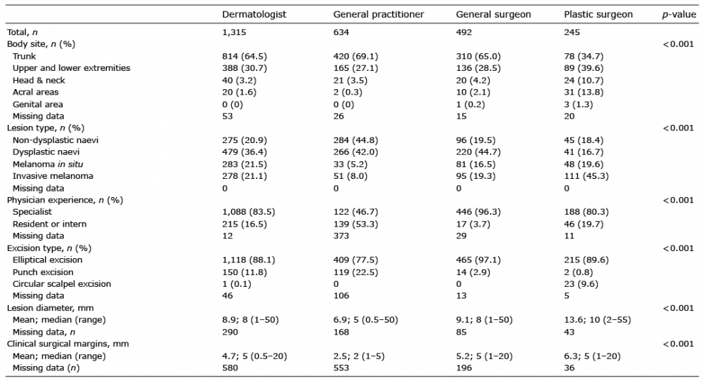 Click to show fullsize
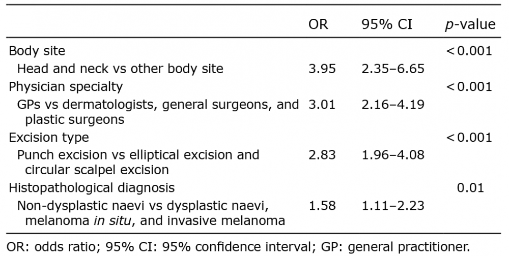 Click to show fullsize