


1Department of Dermatology, Hospital of Västmanland Västerås, Västerås, Sweden, 2Department of Dermatology, Zealand University Hospital, Roskilde, 3Department of Clinical Medicine, Faculty of Health Sciences, University of Copenhagen, Copenhagen, Denmark, 4Department of Dermatology and Venereology, Institute of Clinical Sciences, Sahlgrenska Academy, University of Gothenburg and 5Region Västra Götaland, Sahlgrenska University Hospital, Department of Dermatology and Venereology, Gothenburg, Sweden. E-mail: sara.calander@regionvastmanland.se
Accepted Aug 25, 2021; Epub ahead of print Aug 26, 2021
Acta Derm Venereol 2021; 101: adv00551.
doi: 10.2340/00015555-3909
Tinea capitis (TC) predominantly affects prepubertal children (1, 2). During the 20th century the prevalence of TC in western society increased due to increasing migration and travel. The causative pathogens in TC are changing over time and geographical area (3, 4). In the early 20th century Microsporum audouinii was one of the most prevalent causative agents of TC in Europe. This changed with the introduction and widespread use of griseofulvin and, after the 1950s, other dermatophyte species became dominant pathogens in TC (5).
In the Nordic countries, Trichophyton species are the most common causative pathogen in TC, and oral terbinafine is considered first-line treatment (6–8). Griseofulvin is generally considered the most efficient treatment for Microsporum species, but is difficult to obtain in Sweden as it requires a specific license application for prescription, often resulting in delay in starting treatment.
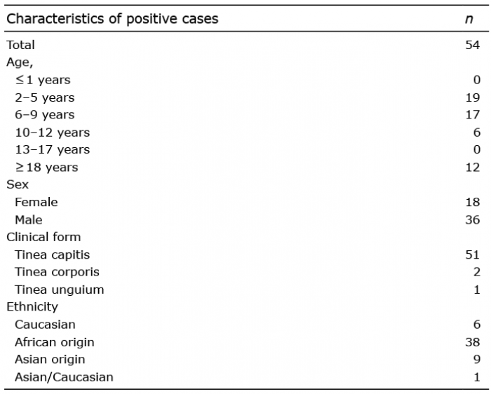
In 2019 an outbreak of TC caused by M. audouinii started in central Sweden (in Västmanland County). It took more than one year to control the outbreak. A total of 54 children (median age 6 (range 2–12) years were found to be positive for M. audouinii during this period (Tables I, II, and Tables SI and SII). Given the extent of the outbreak, we report here 2 representative case reports, which may provide useful insights for future outbreaks.
Table I. Distribution of positive cases of tinea capitis caused by Microsporum audouinii
Table II. Patients on systemic therapy: treatment effect
In the Department of Dermatology, Västerås, patients are typically referred from general practitioners and some already have a culture-proven infection. All family members are examined for tinea by a dermatologist and a nurse. Wood’s light is used for infection screening and to find the optimal location for hair plucking. The material is sent to the microbiology laboratory for direct microscopy and culture. Asymptomatic individuals are screened using a small brush, which is rubbed on the scalp and then cultured in Sabouraud glucose agar. In addition to oral antifungal therapy, ketoconazole shampoo is used as an adjuvant therapy to prevent spread of the disease, and hygienic measures, such as disinfection of brushes, combs and cutting tools and washing hair-pieces at 60°C, is recommended.
Case 1. A 3-year-old boy, who was attending a day-care centre, presented at the Department of Dermatology, Västerås. The patient had widespread skin scaling of the scalp, diffuse alopecia and 3 red scaly plaques on his trunk. Fungal cultures showed growth of M. audouinii from the trunk and Trichophyton violaceum from the scalp. Oral terbinafine according to weight (62.5 mg once daily) and ketoconazole shampoo (twice weekly) were prescribed for 6 weeks. The boy had no siblings and culture screening of the parents were negative. At 4 weeks follow-up the scalp had improved, with just a few residual scaly patches, and the infection of the trunk had cleared. Sixteen weeks later scalp samples were obtained and demonstrated growth of M. audouinii. Oral terbinafine was reinitiated and prescribed for 4 weeks . The asymptomatic parents were re-screened, and oral terbinafine was initiated due to growth of M. audouinii. The index-boy presented with widespread scaly patches on the scalp at 10 weeks follow-up (22 weeks after the initial presentation). Cultures still showed growth of M. audouinii, and oral terbinafine was reinitiated. A new culture after 4 weeks was still positive, and treatment was switched to griseofulvin microsize (20 mg/kg body weight) daily for 6 weeks, which eventually resulted in clearance of symptoms and negative mycological cultures at 8 weeks follow-up. The M. audouinii in the parents also cleared on griseofulvin.
Case 2. A 5-year-old boy presented with an itchy discretely scaly patch with hair loss on the posterior aspect of the scalp (Fig. 1). The patient attended a day-care centre that was affected by the outbreak. The parents and 3 siblings were examined by culture, and a 6-year-old sister and a 7-year-old brother presented with symptoms suggestive of TC, which were confirmed by growth of M. audouinii. The index patient was treated orally with terbinafine 125 mg once daily for 6 weeks. The symptoms worsened during the course of treatment and therefore he was scheduled for a new appointment after 4 weeks. The terbinafine treatment was prolonged 6 weeks and the dose doubled 2 weeks later due to disease progression for a further 4 weeks, but, as the symptoms persisted, the treatment was switched to oral fluconazole (6 mg/kg body weight daily). At follow-up, 4 weeks later, there were no remaining symptoms and the fungal culture was negative. The 7-year-old brother and 6-year-old sister were treated similarly, but the sister did not heal as quickly and needed an extended fluconazole treatment before her TC cleared. A few months after clearing of the lesions, the family was again referred with a new-born baby. This time a 2-year old boy in the family was the index patient, with TC caused by M. audouinii. Mycological screening of the other family members was performed and all were negative. The index patient was treated successfully with griseofulvin microsize (20 mg/kg body weight) for 8 weeks.
Fig. 1. Scalp of a patient with tinea capitis caused by Microsporum audouinii. A round patch is visible, with broken hairs and discrete skin scaling.
Our experience from this outbreak supports the use of griseofulvin as the first-line drug for M. audouinii infection, which is in agreement with the latest Cochrane Review on treatment of TC (6). Nonetheless, this review is based on only 2 investigations, in which the majority of Microsporum infections were caused by M. canis and not M. audouinii (9, 10). The outbreak abated only after griseofulvin was initiated as first-line treatment. In the prepubertal children, in the current outbreak, more boys than girls were infected, which is consistent with previous reports (1, 2, 4, 5). Possible contributing factors to be considered are the boys’ short haircuts, which increase access to the scalp roots, as well as increasing the risk of infection via sharing of hair trimmers and the chance of detecting symptoms (11–13).
The infection control unit was involved early and made a survey. They identified selected day-care centres as important sources of disease transmission and provided information to the staff and parents. No screening of all children at the affected institutions was performed. Infection control staff visited affected day-care centres and found hairdresser corners in which combs and hair accessories were shared without cleaning. Moreover, children shared mattresses during rest. Routines pertaining to individual mattress protectors and cabinets for storing were introduced. After the outbreak, the dermatologists at our department have become more thorough, keeping a record of which day-care centre the child attends, in order to detect the spread of infection and involve the infection control unit. Guidelines have been considered regarding testing all children at the same time, if possible, in an affected day-care centre, even asymptomatic children.
Few smaller outbreaks of M. audouinii have been described previously in Munich and Switzerland (14, 15). In comparison with the outbreak described in this article they were managed differently, mainly through infection control measures. For example, in Switzerland (with an outbreak of only 3 individuals) all children attending the same school classes were screened. In Munich the children were not allowed to return to school until 3 consecutive cultures were negative.
Despite a shift to a successful treatment regime and support from the infection control unit, sporadic cases of TC caused by M. audouinii are still being referred to our department. Importantly, we have not yet observed any cases in which treatment with griseofulvin has been unsuccessful, which supports its usefulness in treating this infection.
The authors have no conflicts of interest to declare.


 Click to show fullsize
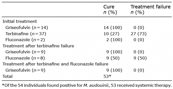 Click to show fullsize
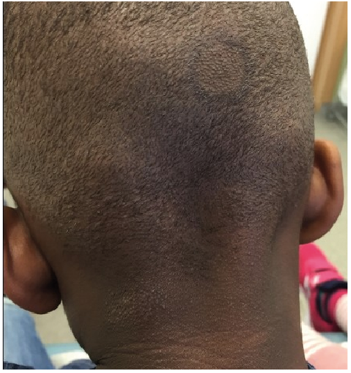 Click to show fullsize