


1Department of Dermatology, Keio University School of Medicine, 35 Shinanomachi, Shinjuku-ku, Tokyo 160-8582, 2Department of Dermatology, National Hospital Organization Tokyo Medical Center, and 3Department of Dermatology, Kyorin University Faculty of Medicine, Tokyo, Japan. *E-mail: akiharu@keio.jp
Accepted Sep 2, 2021; Epub ahead of print Sep 7, 2021
Acta Derm Venereol 2021; 101: adv00549.
doi: 10.2340/00015555-3920
Spirally twisted rolled hairs have been reported in association with follicular hyperkeratotic conditions, including keratosis pilaris and ichthyosis (1). Rolled hairs appear mostly on the extensor extremities and trunk, but have not been reported on the scalp (1). Naevus comedonicus (NC) is a rare naevus caused by somatic NEK9 mutations, which shows multiple comedo formation within the affected skin (2). We report here 2 cases of NC of the scalp presenting with rolled hairs, detected by trichoscopy.
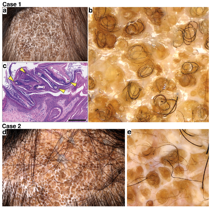
Case 1. Case 1 was a 1-year-old Japanese girl with a congenital naevus on the parietal region of the scalp. The patient was otherwise healthy and had an unremarkable family history. The naevus presented a cluster of hyperkeratotic, brownish papules with alopecia, showing a cobblestone appearance (Fig. 1a). Trichoscopic examination revealed heavily rolled hairs within papules (Fig. 1b). Skin biopsy of the lesion revealed rolled hairs embedded in follicular plugs and a dilated hair follicle infundibulum (Fig. 1c).
Fig. 1. Clinical, trichoscopic, and histopathological features of the patients. The features of case 1 and case 2 are shown in upper panels (a–c) and lower panels (d–e), respectively. (a, d) A cluster of hyperkeratotic, brownish papules with alopecia on the scalps. (b, e) Trichoscopic examination of the patient’s scalp showing rolled hairs within papules. (c) Rolled hairs (yellow arrowheads) embedded in follicular plugs with a dilated hair follicle infundibulum (haematoxylin-eosin; scale bars, 250 µm).
After obtaining written informed consent and approval from the Institutional Review Board, genetic analyses of genomic DNA isolated from peripheral blood and the lesional epidermis and dermis, which were separated by dispase treatment, were performed (3). Somatic heterozygous mutations in NEK9 were identified; c.1718T>C (p.I573T) in case 1 and c.1755_1757delAAC (p.T586del) in case 2, in an epidermis-specific manner (Fig. 2). The somatic mutations of NEK9 identified in the current cases have been previously reported in NC (2, 4). The diagnosis of NC was therefore confirmed in both cases.
Fig. 2. Analysis of NEK9 in genomic DNA isolated from the biopsied skin and peripheral blood leukocytes. Sanger sequencing chromatograms of cases 1 and 2 are shown in upper panels and lower panels, respectively. Arrows indicate the mutations of NEK9 identified in an epidermis-specific manner.
NC is an uncommon malformation of the pilosebaceous unit characterized by dilated follicular orifices filled with follicular plugs. In NC, massive cystic inflammation and fibrosis sometimes occur during adolescence (5), as happened in case 2.
NC is most commonly located on the face, neck, trunk and arms. While several cases of NC have been reported to develop on the scalp, trichoscopic findings were described for only one adult patient with follicular plugs and vellus hair (6). To the best of our knowledge, this is the first report describing a unique trichoscopic finding of NC of the scalp. Both case 1 and case 2 exhibited rolled hairs of the scalp embedded within follicular plugs. The waxy follicular plugs caused by abnormal keratinization were speculated to have hindered the emergence of hair, which resulted in the spiral transverse hair growth within the semilucent structures, probably via slower growth of follicular plugs than hairs (7). Most of the lesional hairs were rolled hairs in case 1, which could be associated with the softness of babyhood hairs. The lesional hairs of case 1 were considered not hard enough to penetrate the follicular plugs.
It is noteworthy that differential diagnosis of NC of the scalp includes naevus sebaceous, within which various neoplasms can occur (8). Cerebriform patterns and yellowish globules in cobblestone patterns are unique trichoscopic features of naevus sebaceous in adulthood and childhood, respectively (9). Our observation suggested that rolled hairs within follicular plugs are potential diagnostic clues for NC of the scalp, which can be useful for differentiation from naevus sebaceous. Further accumulation of cases with NC of the scalp would enable the establishment of its trichoscopic features.
The patients in this manuscript have given written informed consent to publish their case details.
Conflicts of interest. M. Ohyama is a scientific advisor for Eli Lilly Japan, Pfizer Japan Inc., Taisho Pharmaceutical Co., and RHOTO Pharmaceutical Co. and received research grants not related to this study from Shiseido Co. The other authors have no conflicts of interest to declare.


 Click to show fullsize
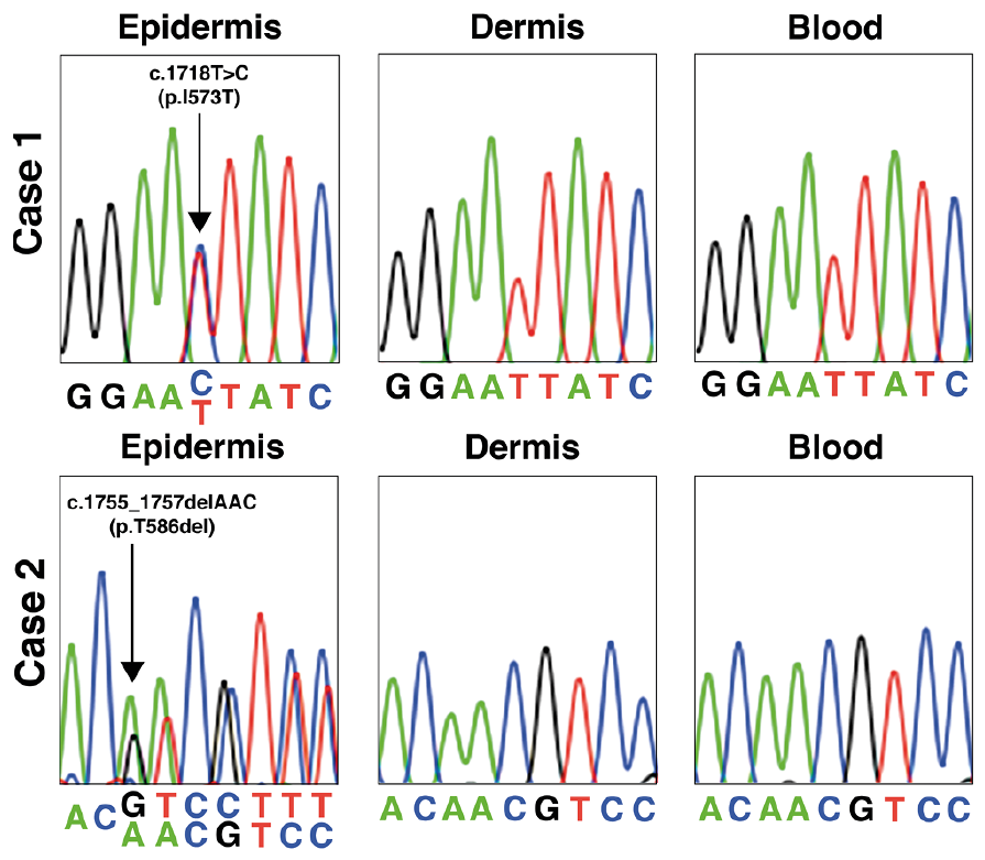 Click to show fullsize