Birgit Skoffer, PhD1,2, Ulrik Dalgas, PhD3, Inger Mechlenburg, PhD4, Kjeld Søballe, DMSc4 and Thomas Maribo, PhD5,6
From the 1Health, Aarhus University, 2Department of Physical and Occupational Therapy, Aarhus University Hospital, 3Department of Public Health, Section of Sport Science, Aarhus University, 4Orthopaedic Research Centre, Aarhus University Hospital, 5Rehabilitation Center Marselisborg, Department of Public Health, Section of Social Medicine and Rehabilitation, Aarhus University and 6Public Health and Quality Improvement, Central Denmark Region, Aarhus, Denmark
OBJECTIVE: To determine whether muscle strength in patients scheduled for total knee arthroplasty is: (i) strongly associated with both measured functional performance and patient-reported measures; (ii) more closely associated with functional performance when measured during concentric than during isometric contractions; and (iii) more strongly related to the 30-s chair stand test than to the timed-up-and-go and walking measures.
DESIGN: Cross-sectional-study.
Patients: Fifty-nine patients (36 women, 23 men), mean age 70.4 years.
METHODS: Associations between muscle strength, measured functional performance, and patient-reported measures were calculated.
RESULTS: Both knee extensor and knee flexor strength were associated with performance-based measures. In general, concentric knee flexor muscle strength was more strongly associated with functional performance than was isometric knee flexor strength. Concentric and isometric knee extensor strength were of equal importance. The 30-s chair stand test was better than the timed-up-and-go and the walking tests at determining muscle strength.
CONCLUSION: Future rehabilitation programmes should include both the knee extensor muscles and the knee flexor muscles in order to improve functional performance. The 30-s chair stand test is a valid and clinical relevant proxy measure of knee extensor and knee flexor muscle strength.
Key words: osteoarthritis; knee; muscle strength; functional capacity.
J Rehabil Med 2015; 47: 00–00
Correspondence address: Birgit Skoffer, Department of Physical and Occupational Therapy, Aarhus University Hospital, 8000 Aarhus, Denmark. E-mail: bss@mph.au.dk
Accepted Nov 5, 2014; Epub ahead of print Feb 12, 2015
INTRODUCTION
Knee osteoarthritis (OA) is a frequent health problem in older adults (1). The most prominent symptoms of knee OA are pain, reduced functional performance, decreased muscular strength and reduced quality of life (2–5). Pain and reduced functional performance in combination with radiographically confirmed severe OA are the main indications for total knee arthroplasty (TKA) (6).
Impairment of knee extensor muscle strength in patients with knee OA is well-documented (3), and decreased knee extensor muscle strength is the only significant determinant of reduced functional performance in patients with severe knee OA (7). Furthermore, knee extensor muscle weakness is a primary risk factor for developing knee pain, disability and progression of joint damage in persons with knee OA (8, 9). Although focus in the literature is largely on the knee extensors, substantial knee flexor strength deficits have been reported (3). Furthermore, concentric knee flexor strength is a significant predictor of the performance of a stair-climbing task (3), and isometric knee flexor strength significantly correlates with stair climbing, standing from sitting, walking, and lifting an object from the floor (3). The importance of knee muscle strength is evident, but valid and reliable tests of muscle strength require expensive and complex laboratory equipment. If it were possible to identify a functional performance test that is closely associated with muscle strength, such a test could be used as a surrogate for muscle testing, e.g. dynamometry testing.
Patients awaiting TKA perform more poorly in performance-based measures, such as the timed-up-and-go (TUG) test, the 6-min walk test (6MWT), and the single-limb stance test (10) compared with healthy controls. However, knowledge about how these performance-based measures relate to knee muscle strength is sparse. This knowledge is important for clinicians who monitor the results of a rehabilitation intervention with performance-based measures in patients with knee OA in a clinical setting and who need to know which performance-based measure is to be preferred.
Most studies on knee muscle strength are based on isometric muscle tests (3). Isometric tests of the muscle groups may reflect their static positions, whereas the results of concentric muscle tests probably better reveal the complex activation of muscle groups exhibited in physical function. Knowledge of the association between isometric and concentric muscle strength and performance-based measures may help to determine the best clinically relevant method for testing knee muscle strength in the laboratory. Moreover, a better understanding of the relationship between knee extensor and knee flexor muscle strength and performance-based measures may improve rehabilitation programmes before and after TKA (11, 12).
The aim of this study was to test the hypotheses that, in patients scheduled for TKA, knee extensor and knee flexor strength would be: (i) weaker in the affected leg than in the non-affected leg; (ii) strongly associated with both measured functional performance and patient-reported measures; (iii) more closely associated with functional performance when measured during concentric than during isometric contractions; and (iv) more strongly related to the 30-s chair stand test (30sCST) than to the TUG test and walking measures.
METHODS
Study design and patients
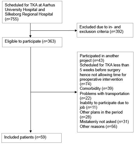
This cross-sectional study is part of a randomized, controlled study that investigates the effect of preoperative progressive resistance training on functional performance and muscle strength after TKA. A total of 59 patients scheduled for TKA were included from the Orthopaedic Department of Aarhus University Hospital and Silkeborg Regional Hospital, Denmark (Fig. 1).
Included were patients who were: (i) scheduled for primary unilateral TKA; (ii) diagnosed with OA; (iii) resident in the Aarhus municipality; (iv) able to transport them-selves to training; and (v) willing to provide informed consent. Excluded were patients who were: (i) age < 18 years; (ii) had heart disease or uncontrolled hypertension; (iii) had neuromuscular or neurodegenerative conditions; and (iv) were unable to comprehend the protocol instructions.
The study followed the Declaration of Helsinki, was approved by the regional Ethics Committee (journal no. M-20110191) and was registered with the Danish Data Protection Agency (registration no. 1-16-02-191-11) and at ClinicalTrials.gov (NCT01647243).
Test procedures
The assessment included tests of muscle strength and functional performance and measurement of height, body mass and range of knee joint motion. Furthermore, patients completed questionnaire items on pain, functional performance and quality of life. All patients were tested according to the protocol 6 weeks before TKA by the same assessor (BS).
Muscle strength. Muscle strength was measured using an isokinetic dynamometer (Humac Norm, Computer Sports Medicine Inc., Massachusetts, USA). Patients were in a seated position with a 90° hip flexion. The body and the tested thigh were fastened with straps. The anatomical axis of the knee was aligned with the axis of the dynamometer, and the ankle cuff was 3 cm proximal to the medial malleolus. Moment values were corrected for the gravity of the lower limb and were measured by the dynamometer at a knee joint angle of 45°.
Patients performed 3 maximal isometric contractions of the knee extensors at a knee joint angle of 70° (0° = full knee extension) and of the knee flexors at a knee joint angle of 20° (13). Rest periods of 60 s were allowed between attempts. The trial with the highest peak torque (Nm) was selected for further analysis. Isometric testing was performed bilaterally.
The concentric knee extensor and knee flexor muscle strength of the affected knee was evaluated at 60°/s (peak moment, Nm). The patients performed 6 maximal concentric contractions with full possible range of motion (ROM); the trial with the highest peak torque was selected for further analysis.
Dynamometry is considered the gold standard of muscle strength assessment, and dynamometry tests of knee extensor muscles in knee OA have proven reliable (14).
The 30sCST measures the total number of full rises to standing position that patients were able to perform in 30 s. Patients were seated in a standard chair with their arms folded across their chest (15). The best of 2 test trials was selected for further analysis. The test is reliable in patients with knee OA (16, 17).
TUG measures (in s) the time taken to rise from a standard armchair, walk 3 m, turn, walk back to the chair and sit down again. Patients were instructed to walk as fast as they felt was safe, and the use of an assistive device was allowed if necessary (18). The fastest time of 2 test trials was selected for further analysis. The test is valid in patients with knee OA (16, 19).
Ten-metre walk test (10mWT). This test measures maximal walking speed. Patients were instructed to walk 12 m between 2 marked lines. The timer was stopped when the first foot touched or passed the 10-m line. Patients were instructed to walk as fast as they felt was safe using an assistive device if necessary (20). The fastest time of 2 test trials was selected for further analysis.
Six-min walk test (6MWT). This test measures maximal walking distance in 6 min. Subjects were instructed to walk as far as possible in 6 min in a safe manner in an undisturbed 30-m long corridor. The use of assistive devices was allowed if necessary (21). The test is reliable in patients with knee OA (22).
Active and passive knee joint flexion and the extension ROM of the affected knee were measured by goniometry. The patient was placed in the supine position. The goniometer fulcrum was placed over the lateral epicondyle with one 30-cm arm pointed towards the major trochanter of the femur and the other towards the lateral malleolus (23). During active ROM, patients flexed and extended the knee as much as possible. During passive ROM, the assessor flexed and extended the knee until the patient said “stop”. The method is reliable and valid in patients with knee restrictions (23).
Knee injury and Osteoarthritis Outcome Score (KOOS). This patient-reported questionnaire consists of 5 subscales: pain, other symptoms, function of daily living, function in sport and recreation, and knee-related quality of life (24). The KOOS is a reliable and valid tool in patients with knee OA and TKA (25–27). However, in sport and recreation the subscale function has shown weak-to-moderate reliability and weak construct validity (25, 26).
Knee pain ratings. Ratings were recorded on an 11-point numerical rating scale from 0 (“no pain”) to 10 (“worst pain imaginable”). Current pain, the worst pain during the past 14 days, and average pain during the past 14 days were rated. The numerical rating scale is a reliable and valid tool for pain assessment (28).
Statistical analyses
Descriptive statistics were calculated, using mean and standard deviation (SD) for normally distributed data and median and range if data showed non-normal distribution. Normal distribution of data was checked with box-plots, q-q plots, histograms and dot-plots. To compare muscle strength between the affected and the non-affected leg, paired t-test was applied. To calculate the association between functional performance, patient-reported outcomes and knee muscle strength, linear regression analyses were applied. Logarithmic transformation was applied on the non-normally distributed data to achieve an approximate, normal distribution. Pitman’s test was applied to identify which functional performance test had the closest relationship with muscle strength and whether concentric or isometric strength had the closest relationship with functional performance. Statistical analyses were performed in Stata version 12.1 (StataCorp LP, USA).
RESULTS
A total of 59 patients agreed to participate in the study during the inclusion period (Fig. 1). Their demographics are shown in Table I. Median values and range for functional performance tests and muscle strength are shown in Table II.
|
Table I. Characteristics of included patients scheduled for total knee arthroplasty |
|
|
Patients’ characteristics |
|
|
Sex, female/male, n |
36/23 |
|
Age, years, mean (SD) |
70.4 (6.8) |
|
Height, m, median (range) |
1.68 (1.45–1.97) |
|
Body mass, kg, median (range) |
84.0 (56.8–137.4) |
|
Body mass index, kg/m2, median (range) |
30.3 (22.6–42.5) |
|
Range of motion, mean (SD) |
|
|
Knee flexion AROM (°) |
119.7 (10.1) |
|
Knee flexion PROM (°) |
124.2 (9.7) |
|
Knee extension AROM (°)a |
5.9 (3.1) |
|
Knee extension PROM (°)a |
4.1 (3.4) |
|
Patient-reported outcomes |
|
|
KOOS pain (median (range) |
50.0 (27.8–88.9) |
|
KOOS other symptoms (median (range) |
53.6 (21.4–96.4) |
|
KOOS function of daily living (median (range) |
55.9 (29.4–88.2) |
|
KOOS sport & recreation (median (range) |
20.0 (00.0–75.0) |
|
KOOS quality of life (median (range) |
37.5 (6.3–81.3) |
|
Current painb, median (range) |
5 (1–10) |
|
Worst pain during the past 14 daysb, median (range) |
7 (3–10) |
|
Average pain during the past 14 daysb, median (range) |
5 (1–10) |
|
aLack of full extension. bMeasured on 11-point numerical rating scale. KOOS: Knee injury and Osteoarthritis Outcome Score; AROM: active range of motion; PROM: passive range of motion; SD: standard deviation. |
|
Muscle strength in affected and non-affected leg
The median values and the range of concentric and isometric knee muscle strength are shown in Table II. The knee extensors were significantly weaker in the affected leg than in the non-affected leg (p < 0.01), whereas for knee flexors the difference between the 2 legs was insignificant (p = 0.51). The mean strength of the knee extensors in the affected leg corresponded to 89.1% (SD 30.2) of that of the non-affected leg.
|
Table II. Functional performance and knee extension and flexion muscle strength in included patients |
|
|
Median (range) |
|
|
Functional performance 30sCST (rep) TUG (s) 10mWT (s) 6MWT (m) Normalized muscle strength Affected leg Concentric extension peak torque (Nm/kg) Concentric flexion peak torque (Nm/kg) Isometric extension peak torque (Nm/kg) Isometric flexion peak torque (Nm/kg) Non-affected leg Isometric extension peak torque (Nm/kg) Isometric flexion peak torque (Nm/kg) |
11 (0–23) 8.6 (5.6–21.2) 7.6 (4.8–13.6) 420 (120–592) 0.9 (0.3–1.8) 0.4 (0.1–1.3) 1.0 (0.3–1.7) 0.6 (0.2–1.8) 1.1 (0.5–2.6) 0.5 (0.3–1.4) |
|
30sCST: 30-s chair stand test; TUG: timed-up-and-go; 10mWT: 10-m walk test; 6MWT: 6-min walk test; rep: repetitions. |
|
Muscle strength vs functional performance/patient-reported measures
An overall association was found between functional performance and concentric and isometric knee extensor and knee flexor muscle strength in the affected and non-affected leg, except for the 6MWT. The association was generally strongest for the affected leg (Table III).
|
Table III. Associations between functional performance measures and muscle strengtha |
|||||||||||
|
Muscle strength |
CST (rep.)b |
|
TUG (s)b |
10mWT (s)b |
6MWT (m)b |
||||||
|
Crude β (p) |
Adjustedc β (p) |
|
Crude β (p) |
Adjustedc β (p) |
Crude β (p) |
Adjustedc β (p) |
Crude β (p) |
Adjustedc β (p) |
|||
|
Affected leg |
|||||||||||
|
Concentric extension peak torque, Nmb |
0.29 (0.01) |
0.49 (< 0.01) |
–0.23 (< 0.01) |
–0.26 (< 0.01) |
–0.17 (< 0.01) |
–0.18 (0.01) |
0.17 (0.03) |
0.13 (0.18) |
|||
|
Concentric flexion peak torque (Nm)b |
0.28 (< 0.01) |
0.32 (< 0.01) |
–0.21 (< 0.01) |
–0.18 (0.01) |
–0.17 (< 0.01) |
–0.16 (< 0.01) |
0.20 (0.01) |
0.16 (0.02) |
|||
|
Isometric extension peak torque (Nm)b |
0.28 (0.05) |
0.58 (< 0.01) |
–0.21 (< 0.01) |
–0.21 (0.06) |
–0.17 (< 0.01) |
–0.19 (0.02) |
0.18 (0.03) |
0.19 (0.09) |
|||
|
Isometric flexion peak torque (Nm)b |
0.28 (0.02) |
0.43 (< 0.01) |
–0.14 (0.09) |
–0.04 (0.73) |
–0.12 (0.05) |
–0.06 (0.46) |
0.14 (0.12) |
0.00 (0.97) |
|||
|
Non-affected leg |
|||||||||||
|
Isometric extension peak torque (Nm)b |
0.23 (0.08) |
0.55 (< 0.01) |
–0.24 (< 0.01) |
–0.27 (0.03) |
–0.16 (< 0.01) |
–0.17 (0.07) |
0.14 (0.10) |
0.06 (0.65) |
|||
|
Isometric flexion peak torque (Nm)b |
0.27 (0.06) |
0.34 (0.06) |
–0.21 (0.02) |
–0.16 (0.19) |
–0.15 (0.03) |
–0.10 (0.25) |
0.21 (0.03) |
0.15 (0.21) |
|||
|
aAnalysed by linear regression; bLog-transformed data; cAdjusted for age, sex, height, and weight; β, Regression coefficient. 30sCST: 30-s chair stand test; TUG: timed-up-and-go; 10mWT: 10-m walk test; 6MWT: 6-min walk test; Rep: repetitions. |
|||||||||||
Scores of the KOOS subscales are shown in Table I. Using linear regression, we found no association between subscales and any knee muscle strength parameters, either for crude or for adjusted scores (Table SI1). In contrast, an overall association was found between the KOOS subscales and pain (Table SII1).
Concentric vs isometric muscle strength
In general, both concentric and isometric knee extensor and knee flexor muscle strength were associated with functional performance-based measures. However, concentric knee flexor strength was more closely associated with the TUG, 10mWT and the 6MWT than isometric knee flexor strength, but no difference was found between concentric and isometric knee extensor strength in any test of functional performance (Table IV).
|
Table IV. Comparison of associations between functional performance and muscle strength measures of the affected leg; concentric vs isometric |
||||
|
30sCST |
TUG |
10mWT |
6MWT |
|
|
Knee extension peak torque, Nm |
NS |
NS |
NS |
– |
|
Knee flexion peak torque, Nm |
NS |
concentric > isometric p < 0.01 |
concentric > isometric p < 0.01 |
concentric > isometric p = 0.04 |
|
*Analysed by Pitman’s test. 30sCST: 30-s chair stand test; TUG: timed-up-and-go; 10mWT: 10-m walk test; 6MWT: 6-min walk test; NS: non-significant; >: indicates stronger association; <: indicates weaker association. |
||||
30sCST vs TUG and walking
The 30sCST was the test most strongly associated with all parameters of muscle strength. The 30sCST was more closely associated with both concentric and isometric knee extensor and knee flexor than the TUG and the walking tests (Table V).
|
Table V. Comparison of associations between functional performance and muscle strength measures |
|||||
|
CST vs TUG |
CST vs 10mWT |
CST vs 6MWT |
TUG vs 10mWT |
10mWT vs 6MWT |
|
|
Affected leg |
|||||
|
Concentric knee extension peak torque, Nm |
CST > TUG p < 0.01 |
CST > 10mWT p < 0.01 |
CST > 6MWT p < 0.01 |
TUG > 10mWT p < 0.01 |
10mWT > 6MWT p < 0.01 |
|
Concentric knee flexion peak torque, Nm |
CST > TUG p < 0.01 |
CST > 10mWT p < 0.01 |
CST > 6MWT p < 0.01 |
TUG < 10mWT p < 0.01 |
10mWT > 6MWT p < 0.01 |
|
Isometric knee extension peak torque, Nm |
CST > TUG p < 0.01 |
CST > 10mWT p < 0.01 |
CST > 6MWT p < 0.01 |
TUG < 10mWT p < 0.01 |
10mWT > 6MWT p < 0.01 |
|
Isometric knee flexion peak torque, Nm |
CST > TUG p < 0.01 |
CST > 10mWT p < 0.01 |
CST > 6MWT p < 0.01 |
– |
– |
|
Non-affected leg |
|||||
|
Isometric knee extension peak torque, Nm |
CST > TUG p < 0.01 |
CST > 10mWT p < 0.01 |
CST > 6MWT p < 0.01 |
TUG > 10mWT p < 0.01 |
– |
|
Isometric knee flexion peak torque, Nm |
– |
– |
– |
– |
– |
|
*Analysed by Pitman’s test. 30sCST: 30-s chair stand test; TUG: timed-up-and-go; 10mWT: 10-m walk test; 6MWT: 6-min walk test; NS: non-significant; >: indicates stronger association; <: indicates weaker association. |
|||||
DISCUSSION
This study shows that knee extensors were weaker in the affected leg than in the non-affected leg, whereas knee flexor muscle strength was similar in the 2 legs. In general, knee extensor and knee flexor muscle strength were associated with performance-based measures, except for the 6MWT. However, no association was observed between patient-reported measures and muscle strength. Concentric muscle strength was generally more closely associated with performance-based measures than isometric muscle strength. Finally, the 30sCST test was the performance-based measures most closely associated with the various parameters of muscle strength.
Muscle strength in affected and non-affected leg
Concentric and isometric knee extensor muscle strength were higher than the corresponding knee flexor strength in both legs. The isometric knee extensor muscle strength was lower in the affected leg than in the non-affected leg, while the muscle strength of the knee flexor was similar in both legs. These results are in accordance with the findings of Stevens-Lapsley et al. (29), who found that isometric knee extensor strength was lower in the affected than in the non-affected leg (21%; p = 0.03), whereas the strength of the knee flexors was similar in both legs in patients scheduled for TKA (p = 0.70). Brown et al. (11) found that the concentric knee extensor and flexor strength of the affected knee was 24–30% lower than the strength of the unaffected leg in patients scheduled for TKA. The latter finding cannot be directly compared with ours, since we did not investigate the concentric strength of the non-affected leg. However, Brown et al.’s finding suggests that knee OA affects concentric muscle strength more than isometric muscle strength.
Muscle strength vs functional performance/patient-reported measures
Overall, the present study revealed associations between performance-based measures and knee extensor and knee flexor muscle strength, except for the 6MWT. The strongest was generally found for the affected leg, but the functional performance was also affected by the muscle strength of the non-affected leg. These results are in agreement with those presented in a study of Mizner et al., which showed an association between functional performance and quadriceps muscle strength in patients scheduled for TKA (12). In their study, a weaker relationship was observed between muscle strength and the TUG compared with the stair-climbing test. Furthermore, Brown et al. demonstrated that knee flexor strength in the involved leg was the best predictor of the 30sCST in patients scheduled for TKA (13). The 6MWT is often used to assess functional performance in different patient groups; for example, in patients before and after TKA (10, 19). We found a positive association only with concentric knee flexor strength. This study found no associations between patient-reported measures and muscle strength. This is in agreement with Brown et al. (11); whereas Kennedy et al. (5) demonstrated low-to-moderate correlation between patient-reported and performance-based measures. Furthermore, other studies have shown that patient-reported measures of knee function are strongly influenced by pain (31), which the results of our study confirmed.
Concentric vs isometric muscle strength
In general, the present study demonstrated a stronger association between performance-based measures and concentric knee flexor muscle strength than between performance-based measures and isometric knee flexor strength. With regard to knee extensors, concentric and the isometric strength seemed to be equally associated with performance-based measures. Concentric knee flexor muscle strength was more strongly associated with the TUG and walking tests than was isometric knee flexor strength.
TUG was closely associated with both concentric and isometric muscle strength of both legs. The TUG was reviewed by the Osteoarthritis Research Society International (OARSI) (16), and is 1 of 5 performance-based measures recommended for research and clinical practice (32).
30sCST vs TUG and walking
The 30sCST was the performance-based measure that most accurately measured muscle strength. Rising from a chair demands considerable strength, and a strong association was expected. Laboratory research of movement analysing kinetic and kinematic parameters demonstrated that the chair stand movement was both selective and showed functional content validity in TKA (31). Furthermore, chair stand movement has been recognized as a biomechanical instrument identifying how knee function is affected (31). In line with our findings, the 30sCST was one of the best rated tests in a review evaluating the properties of performance-based measures to assess physical function in hip and knee OA (16), and 1 of the 3 core tests recommended by the OARSI (32).
Clinical implications
Much attention has been paid to knee extensor muscle strength in clinical research and rehabilitation programmes in clinical practice. However, along with results obtained in other OA patients (33) and healthy controls (3), the results of the present study suggest that it is equally important to include the knee flexor muscles in rehabilitation programmes to improve or maintain functional performance. Furthermore, the 30sCST test was found to be the best proxy measure of muscle strength when more advanced equipment for measurement of knee extensor and knee flexor muscle strength is not available.
Study limitations
As this is a cross-sectional study, we cannot comment on causality, but only on associations between functional performance, patient-reported measures and muscle strength. It may be relevant to investigate the impact of the strength of other muscles in the lower extremity, e.g. the strength of hip and ankle muscles. Furthermore, the sample size did not allow numerous adjustments in the regression analysis.
Conclusion
Future rehabilitation programmes should address both the knee extensor muscles and the knee flexor muscles in order to improve functional performance in patients with knee OA. The 30sCST is a valid and clinical relevant proxy measure of knee extensor and knee flexor muscle strength.
Acknowledgements
The authors acknowledge the following organizations: Aarhus University, Denmark; Department of Physiotherapy and Occupational Therapy, Aarhus University Hospital, Denmark; Bevica Fund, Denmark; Rheumatism Association, Denmark; Danish Physiotherapy Research Fund; Orthopaedic Surgery Research Foundation, Denmark; Aase and Ejnar Danielsen Fund, Denmark; and The Research Foundation of the Central Region Denmark.

1http://www.medicaljournals.se/jrm/content/?doi=10.2340/16501977-1940
References


