


Yoshihiro Kuwano, Hironobu Ihn, Norihito Yazawa, Takashi Kakinuma, Akihiko Asahina, Kanako Kikuchi and Kunihiko Tamaki
Department of Dermatology, University of Tokyo, 7-3-1 Hongo, Bunkyo-ku, Tokyo 113-8655, Japan. E-mail: kuwanoy-tky@umin.ac.jp
Accepted August 3, 2005.
Sir,
Dermatomyositis (DM) is an idiopathic inflammatory myopathy with characteristic cutaneous manifestations: a heliotrope rash, Gottron’s papules and periungual telangiectasia. Cutaneous ulcers are relatively rare. Although systemic corticosteroids combined with other immunosuppressive drugs are frequently effective for the myositis, they are sometimes insufficient to manage the cutaneous manifestations including ulcers. Recently, accumulated evidence has revealed that intravenous immunoglobulin (IVIG) therapy is effective in the management of myositis and cutaneous lesions in DM (1). However, little is known about the efficacy of IVIG on cutaneous ulcers. We report here a case of DM with ulcers that were resistant to oral corticosteroid, but responsive to high-dose IVIG therapy.
CASE REPORT
A 56-year-old Japanese woman presented with a periorbital skin rash in 1998. After 2 months, she had fever of 39°C with myalgia of extremities and erythema on the fingers. She was treated with intravenous dexamethasone sodium phosphate 8 mg once. Although the fever and myalgia improved, her cutaneous lesions remained. When referred to our hospital she had periungual erythema and Gottron’s papules on the knuckles. However, the heliotropic rash and myalgia had disappeared. Laboratory examination showed elevated serum lactate dehydrogenase levels (307 IU/l) and erythrocyte sedimentation rate (52 mm/h), but no elevation of serum creatinine kinase or serum aldolase levels. Antinuclear antibody, anti-Jo1 antibody and anti-cardiolipin antibodies were not detected. The result of electromyography was consistent with the presence of myopathy, while the biopsy specimen of deltoid muscles showed little evidence of myositis. A skin biopsy specimen of her right middle finger demonstrated slight liquefaction of the basal layer, moderate infiltration of lymphocytes, and deposit of mucin in the upper dermis. Extensive examinations showed no existence of occult cancer. She was diagnosed as having DM and was treated initially with oral prednisolone 60 mg daily. Her cutaneous lesions improved and the dosage of prednisolone was decreased to 12.5 mg daily.
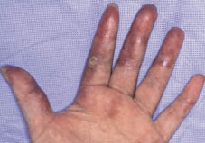
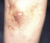
However, approximately 2 years after the initiation of the treatment, the patient began to note dark purple erythema on her fingers, palms, dorsum of hands and elbows, and livedo reticularis on the buttocks. Over the next 2 months, punched-out cutaneous ulcers with induration, as well as necropathy of her right little finger, appeared on the dark purple erythema (Figs 1 and 2). Although she was treated with oral prednisolone up to 20 mg daily in combination with intravenous alprostadil and topical treatment, these ulcers progressed in number and size with local pain. Therefore high-dose IVIG treatment (400 mg/kg daily for 5 days) was initiated. She received IVIG treatment twice. Two days after the initiation, both the pain and induration of ulcers improved dramatically. In addition, the ulcers on her buttocks disappeared and dark purple erythema became faint within a week. Finally a month after the second IVIG infusion, erythema disappeared except on the fingers and the cutaneous ulcers healed. No side effect was noted during the treatment and there has been no relapse for 1 year after the treatment.
Fig. 1. Dark purple erythema on the fingers and palms, and punched-out- shaped cutaneous ulcer of the left forefinger.
Fig. 2. Dark purple erythema and punched-out-shaped cutaneous ulcer with induration on the right elbow.
DISCUSSION
Cutaneous ulcer is seen in about 10% of patients with DM. Although oral corticosteroids combined with other immunosuppressive drugs are frequently used for the treatment of DM (1, 2), these agents are sometimes insufficient to manage all symptoms, especially cutaneous manifestations (2).
IVIG therapy is a very effective treatment for many autoimmune diseases. Several mechanisms for its efficacy have been proposed (3, 4).
Previous studies have shown IVIG therapy to be effective in 33–100% of patients with systemic lupus erythematosus (SLE) (5–7). The improved symptoms and signs are nephritis, thrombocytopenia, encephalitis, leukocytopenia, arthritis, myalgia, fever, asthenia and augmented anti-dsDNA levels. There are two reports about skin lesions in patients with SLE. Schroeder et al. (6) reported that IVIG improved erythema, but De Pita et al. (7) reported no significant modification of skin lesions. As for systemic sclerosis, there is a report of three patients whose total skin thickness score improved after IVIG therapy (8).
Recently, it was also reported that high-dose IVIG therapy results in successive effect on the myopathy of DM (1, 9). In particular, Dalakas et al. (1) reported a controlled study of 15 patients that demonstrated the effectiveness of IVIG therapy for DM. However, few papers have reported on the effect of IVIG therapy on the cutaneous manifestations, especially cutaneous ulcers in DM (1, 10).
Our patient suffered from cutaneous ulcers that were resistant to oral corticosteroid, alprostadil and topical treatment for over 1 year. The cutaneous ulcers immediately healed with IVIG therapy and we observed no recurrence for 1 year after IVIG administration.
REFERENCES
1. Dalakas MC, Illa I, Dambrosia JM, Soueidan SA, Stein DP, Otero C, et al. A controlled trial of high-dose intravenous immune globulin infusions as treatment for dermatomyositis. N Engl J Med 1993; 329: 1993–2000.
2. Callen JP. Dermatomyositis. Lancet 2000; 355: 53–57.
3. Jolles S, Hughes J, Whittaker S. Dermatological uses of high-dose intravenous immunoglobulin. Arch Dermatol 1998; 134: 80–86.
4. Kazatchine MD, Kaveri SV. Immunomodulation of autoimmune and inflammatory diseases with intravenous immune globulin. N Engl J Med 2001; 345: 747–755.
5. Levy Y, Sherer Y, Ahmed A, Langevitz P, George J, Fabbrizzi F, et al. A study of 20 SLE patients with intravenous immunoglobulin – clinical and serologic response. Lupus 1999; 8: 705–712.
6. Schroeder JO, Zeuner RA, Euler HH, Loffler H. High dose intravenous immunoglobulins in systemic lupus erythematosus: clinical and serological results of a pilot study. J Rheumatol 1996; 23: 71–75.
7. De Pita O, Bellucci AM, Ruffelli M, Girardelli CR, Puddu P. Intravenous immunoglobulin therapy is not able to efficiently control cutaneous manifestations in patients with lupus erythematosus. Lupus 1997; 6: 415–417.
8. Levy Y, Sherer Y, Langevitz P, Lorber M, Rotman P, Fabrizzi F, et al. Skin score decrease in systemic sclerosis patients treated with intravenous immunoglobulin – a preliminary report. Clin Rheumatol 2000; 19: 207–211.
9. Cherin P, Herson S, Wechsler B, Piette JC, Bletry O, Ziza JM, et al. Intravenous immunoglobulin for polymyositis and dermatomyositis. Lancet 1990; 336: 116.
10. Peake MF, Perkins P, Elston DM, Older SA, Vinson RP. Cutaneous ulcers of refractory adult dermatomyositis responsive to intravenous immunoglobulin. Cutis 1998; 62: 89–93.

