Sir,
Bone marrow transplantation (BMT) is a widely used treatment for a variety of haematological malignancies and for a rapidly expanding list of inflammatory diseases (1). BMT has resulted in improved long-term survival for many of these patients, especially in paediatric cases (2). The most common preparative transplant regimens consist of chemotherapy and total body irradiation (TBI) to suppress the patient’s immune system and eradicate the malignant neoplasm, followed by transplantation of donor bone marrow to restore haematopoietic function (allogenic BMT) (1). Patients receiving allogenic BMT are predisposed to graft-versus-host disease (GvHD), because the donor graft mounts an immunological response against the immunocompromised host (3).
Chronic GvHD is a common late sequela of BMT. Patients with extensive disease require intensive and long-term immune-suppression. Although GvHD may convey beneficial graft-versus-leukaemia/lymphoma effects, it also entails a significant risk of morbidity and mortality, including the risk of developing cancer (4). While the occurrence of skin cancer in paediatric solid organ transplant recipients has been described in depth (5), the reports on non-melanoma skin cancer in bone-marrow transplanted children, particularly with chronic GvHD, are poorly documented.
CASE REPORTS
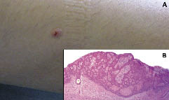
Case 1. An 8-year-old girl was referred to the Dermatology Department from the Department of Paediatrics, University of Pavia, Italy, in 1996 for chronic GvHD. Because of a chemo-resistant acute lymphoblastic leukaemia, she received allogeneic BMT from her HLA-identical father at the age of 4 years, in 1992. The pre-transplant conditioning regimen included TBI (2 Gy × 6), thiotepa (10 mg/kg) and melphalan (140 mg/m2). In order to prevent GvHD, the patient was also treated with cyclosporin A (CyA) (2 mg/kg/day) i.v. and methotrexate (15 mg/m2 day +1, 10 mg/m2 days +3, +6, +11). Eighteen days after BMT the patient developed grade III acute GvHD, with cutaneous, hepatic and intestinal involvement, and was treated with CyA and prednisone. Three months later she developed muco-cutaneous scleroderma-like chronic GvHD, and was treated with topical oral corticosteroids. In the following 5 years, the patient developed a pityriasis rosea-like eruption after hepatitis B vaccination, and hypothyroidism with growth retardation, and was treated with thyroxine and growth hormone. In 2004 the patient developed a dome-shaped, slightly erythematous ulcerated papular lesion, about 1 cm in diameter, on her right leg (Fig. 1a). After surgical excision, histopathology (Fig. 1b) showed proliferation of connecting strands of basaloid cells, with a distinctive basaloid cell layer. The neoplasm was embedded in a prominent fibrous stroma. The histopathological diagnosis was basal cell carcinoma (BCC) with Pinkus’ fibroepithelioma-like features. The patient was seen again in April 2006, with no recurrence.
Fig. 1. Case 1. (a) A flesh-coloured ulcerated sessile papule on the right calf of a 16-year-old girl affected by chronic graft-versus-host disease. (b) Histopathological features of the same lesion: basal cell carcinoma, with Pinkus fibroepithelioma-like features (haematoxylin and eosin).
Case 2. A 14-year-old boy was seen in 1995 for severe chronic GvHD. Because of a chemo-resistant acute myelogenous leukaemia, in 1987 he had received allogeneic BMT from an HLA-identical sister. The pre-transplant conditioning regimen consisted of TBI (2 Gy × 6) and melphalan (140 mg/m2). Methotrexate was also administered for GvHD prophylaxis (15 mg/m2 day +1, 10 mg/m2 days +3, +6, +11). Nine days after BMT the patient developed grade III acute GvHD with cutaneous and intestinal involvement, and was treated with methylprednisone for 60 days. Three weeks after corticosteroid discontinuation, he developed severe chronic GvHD involving the skin, liver and bowel. Therapy included oral prednisone, extracorporeal photopheresis (ECP), azathioprine (AZA) and thalidomide. Scleroderma-like changes were present and involved the trunk and limbs, with mottled appearance, with extensive loss of adnexal structures. Soft tissues, especially on his limbs, were constricted, without pliability. Elbow, knee, and ankle joints were affected by severe functional impairment with flexion contractures. In November 1997, several keratoses, mainly localized on his upper and lower limbs, were successfully treated with cryotherapy. In April 2002, he was seen again for the sudden appearance of an ulcerated and vegetating plaque on the posterior aspect of his left thigh. A 4-mm punch biopsy revealed the histopathological features of a poorly differentiated, infiltrating and ulcerated squamous cell carcinoma (SCC). The lesion was surgically and completely removed. The histopathological diagnosis was poorly differentiated SCC, characterized by a proliferation of strands of poorly differentiated squamous cells, fully involving the dermis. Eight months later the patient had local recurrence, with metastatic involvement of the left inguinal lymph nodes. He died from widespread metastatic disease in May 2003.

Fig. 2. Severe sclerodermoid chronic graft-versus-host disease diffusely involving the skin of patient 2 (age 15 years), causing marked disability. Numerous keratoses, some of them ulcerated, are present.
DISCUSSION
Current information regarding skin cancer in paediatric bone marrow recipients is limited. After 100 days from stem cell transplantation, approximately 50% of patients will experience some degree of chronic GvHD (3). The profound impairment affecting both the cellular and the humoral immunity underlying chronic GvHD is widely documented, as is the strong association between SCC and immunodeficiency (6). Immunosuppressive therapy to control GvHD involves corticosteroids, CyA, AZA, photochemotherapy with oral psoralens and ultraviolet A (PUVA), bath-PUVA, ECP, methotrexate, thalidomide, and, recently, biological response modifiers. All these therapies may be regarded as risk factors for development of cancer. In 1994, Bulengo-Ransby et al. (7) described histopathological bowenoid changes in a patient while receiving CyA to control acute GvHD, only 2 weeks after allogenic BMT.
Nevertheless, to date, very limited reports on the association of GvHD and skin cancer have been published. Recently, Curtis et al. (4) found 183 patients who later developed solid cancers (58 SSCs and 125 non-SCCs) in the course of a follow-up study on 24,011 patients who had undergone haematopoietic stem cell transplantation. In 18 of the 58 patients with SCCs, this lead to death during the follow-up period. The occurrence of SCC and its aggressive behaviour was correlated with the severity of chronic GvHD, long duration of immunosuppressive drug therapy for the treatment of chronic GvHD, in particular the use of AZA. Among the SCC group, 6 patients were younger than 10 years at the time of transplantation, but the authors did not specify data on localization, histopathological features, age at the onset and course in this subset of patients (4).
In the setting of chronic GvHD, the presenting features of SCC may vary. As described by Curtis et al. (4), the neoplasm may be localized either on skin or oral or genital mucosae. Howe & Lang (8), in 1988, described a SCC arising on an ulcer of the left sole in a 20-year-old man with chronic GvHD. The authors considered the plantar ulcer as a feature of the scleroderma-like changes typical of chronic GvHD, predisposing to the occurrence of cancer in that patient. In 1994, Altman & Adler (9) described a 31-year-old man treated with PUVA therapy, oral corticosteroids, CyA, AZA, and thalidomide for chronic GvHD, who developed several hyperkeratotic papules on his arms. Histopathological examinations were consistent with primary well-differentiated SCCs, and 6 subsequent histologically proved primary SCCs on his trunk, arms and legs were excised.
With regard to BCC, its occurrence in children is rare (10). Cases of BCC in the paediatric population have been reported in association with some genodermatoses, such as basal cell nevus syndrome, xeroderma pigmentosum, albinism; superimposed on a nevus sebaceous of Jadassohn; arising on pre-existing scars from burns; in arsenism, or induced by ionizing radiation. Few cases have been reported as being solely related to ultraviolet (UV) radiation and sunburn (11). Ionizing radiation-induced BCCs are not uncommon, but they usually appear in adults after low-voltage superficial radiotherapy for the treatment of benign or malignant skin tumours. The period of latency between exposure to radiation and appearance of BCC varies greatly: average latent period of over 20 years for BCC development has been reported by Garcia-Silva et al. (12). The occurrence of BCCs in children who have been treated with ionizing irradiation for malignancies has been reported in medulloblastomas and basal cell nevus syndrome. BCCs have also been reported in some patients, at an early age, after radiotherapy for acute lymphoblastic leukaemia and internal malignancy (10, 12).
As far the pre-transplant preparation with TBI is concerned, it has independent carcinogenic risks for the development of non-melanoma skin cancer (9). Agents used in the treatment of GvHD in these patients may also increase the carcinogenic risk (4).
The two cases reported here where from a group of 137 pediatric patients with chronic GvHD observed at the Department of Dermatology between 1990 and 2005. Our observations support the view that chronic GvHD may be associated with the occurrence of both SCC and BCC. In particular, patient 1 had a relatively mild course of cutaneous chronic GvHD without ulcerations, and developed BCC 12 years after BMT. She had previously received TBI, thiotepa, melphalan; and later received CyA, methotrexate and oral prednisone. On the other hand, patient 2 had been treated with TBI and melphalan before BMT; prophylaxis for GvHD including, in sequence, methotrexate, methylprednisone, ECP, AZA and thalidomide. He developed invasive SCC 14 years after BMT in the context of severe cutaneous chronic GvHD, with evidence of multiple ulcerations and solar-like keratoses.
The long interval between BMT and the diagnosis of cutaneous malignancy emphasizes the need for careful long-term observation of the cutaneous changes of GvHD.
REFERENCES
1. Robertson KA. Bone marrow transplantation. In: Behrman RE, Kliegman RM, Arvin AM, editors. Nelson textbook of pediatrics. Philadelphia: WB Saunders, 1996: p. 599–609.
2. Locatelli F, Maccario R, Comoli P, Bertolini F, Giorgiani G, Montagna D, et al. Hematopoietic and immune recovery after transplantation of cord blood progenitor cells in children. Bone Marrow Transplantation 1996; 18: 1095 1101.
1101.
3. Horwitz ME, Sullivan KM. Chronic graft-versus-host disease. Blood Rev 2006; 20: 15–27.
4. Curtis RE, Metayer C, Rizzo JD, Socie G, Sobocinski KA, Flowers ME, et al. Impact of chronic GvHD therapy on the development of squamous-cell cancers after hematopoietic stem-cell transplantation: an international case-control study. Blood 2005; 105: 3802–3811.
5. Penn I. De novo malignancy in pediatric organ transplant recipients. Pediatr Transplant 1998; 2: 56–63.
6. Euvrard S, Kanitakis J, Cochat P, Claudy A. Skin cancers following pediatric organ transplantation. Dermatol Surg 2004; 30: 616–621.
7. Bulengo-Ransby SM, Sahn EE, Metcalf JS, Maize JC. Bowenoid change in association with graft-versus-host disease: a cyclosporine toxicity? J Am Acad Dermatol 1994; 31: 1052–1054.
8. Howe NR, Lang PG. Squamous cell carcinoma of the sole in a patient with chronic graft-versus-host disease. Arch Dermatol 1988; 124: 1244–1245.
9. Altman JS, Adler SS. Development of multiple squamous cell carcinomas during PUVA treatment for chronic graft-versus-host disease. J Am Acad Dermatol 1994; 31: 505–507.
10. LeSueur BW, Silvis NG, Hansen RC. Basal cell carcinoma in children. Report of 3 cases. Arch Dermatol 2000; 136: 370–372.
11. Rahbari H, Mehregan AH. Basal cell epithelioma (carcinoma) in children and teenagers. Cancer 1982; 49: 350–353.
12. Garcia-Silva J, Velasco-Benito JA, Pena-Penabad C, Armijo M. Basal cell carcinoma in a girl after cobalt irradiation to the cranium for acute lymphoblastic leucemia: case report and literature review. Pediatr Dermatol 1996; 13: 54–57.