Sumiyuki Mii1,2, Rie Yotsu1, Rika Hayashi1, Harumi Harada1 and Hikaru Eto1
Department of Dermatology, 1St Luke’s International Hospital, Tokyo and 2Kitasato University School of Medicine, 1-15-1 Kitasato, Sagamihara, Kanagawa 228-8555 Japan. E-mail: miisumi@med.kitasato-u.ac.jp
Accepted April 21, 2009.
Sir,
Acquired reactive perforating collagenosis (ARPC) is associated with diabetes mellitus and/or chronic renal failure. The disease lesions consist of many dome-shaped nodules, which have a central umbilication containing firm keratotic plugs. Histologically, transepidermal elimination of degenerated collagen fibres and marked neutrophil infiltrations are characteristic features. ARPC is usually refractory to treatment; however, there have been reports of ARPC treated with phototherapy. In the present case, we treated the patient with narrow-band ultraviolet B (NBUVB) phototherapy. After 20 exposures (total 18.96 J/cm2), all of the nodules had completely disappeared.
CASE REPORT
A 45-year-old woman presented with a 3-month history of skin eruptions without pruritus on her lower extremities. She had had type 2 diabetes, treated with local insulin injections, for 14 years, which was complicated by diabetic nephropathy. The skin eruptions developed on her trunk and extremities. At the time of presentation, there were numerous dome-shaped nodules, which had a central umbilication containing firm keratotic plugs, and erythema was seen around the nodules. There was also a large lesion on the left posterior surface of her leg (Fig. 1A). A skin biopsy was taken from the eruption on her left leg. Histological examination revealed a dome-shaped lesion with a central crater containing parakeratotic horny materials, basophilic granular materials and degenerated collagen. Marked neutrophil infiltration was seen under the dome-shaped lesion. The epidermis demonstrated a cup-shaped depression (Fig. 2A), and Masson’s trichrome staining showed degenerated collagen fibres that had perforated through the epidermis (Fig. 2B). From these findings, a diagnosis of ARPC was made.
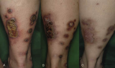
Fig. 1. Clinical course of the left posterior surface of the patient’s leg. (A) Prior to NBUVB irradia=tion. (B) After 10 sessions of irradiation, epidermal regeneration was observed under the keratotic plugs, and resolution of the keratotic plugs left scarring and pigmentation. (C) After 20 sessions of irradiation (total 18.96 J/cm2), all of the nodules completely disappeared.
Fig. 2. Histological features. (A) A dome-shaped lesion with a central crater containing parakeratotic horny materials, basophilic granular materials and degenerated collagen. Marked neutrophil infiltration was seen under the dome-shaped lesion. The epidermis demonstrated a cup-shaped depression (haematoxylin and eosin staining; ×100). (B) Degenerated collagen fibres perforated through the epidermis (Masson’s trichrome staining; ×100).
As treatment with topical corticosteroids worsened the skin eruptions, NBUVB phototherapy was commenced. A fluorescent lamp (TL01) emitting a wavelength of 311 ± 2 nm mounted in a cabinet (UV7001K, Waldmann, Villingen-Schwenningen, Germany), was used. The narrow-band minimal erythema dose (nMED) was 1.2 J/cm2. Irradiation of the whole body surface except for the genital region was commenced at 70% MED (0.8 J/cm2), and the therapeutic dose was increased by 10% at the second irradiation, as per the standard regimen for psoriasis. At the third irradiation (0.96 J/cm2), the erythema decreased, and irradiation was continued at the same dose, 3 times a week. Epidermal regeneration under the keratotic plugs was observed; resolution of the keratotic plugs left behind scarring and pigmentation (Fig. 1B). After 20 exposures (total 18.96 J/cm2), all of the nodules disappeared completely (Fig. 1C). Two years after the treatment, there has been no recurrence.
DISCUSSION
ARPC belongs to the spectrum of primary perforating skin disorders with transepidermal elimination. However, the pathogenesis of ARPC is unknown. In most cases of ARPC, patients experienced severe pruritus and Köbner’s phenomenon. Thus, it is thought that traumatic stimulation, such as scratching, induces the transepidermal elimination of degenerated collagen fibres (1). Recently, there have been reports of ARPC treated with phototherapy including psoralen plus UVA (PUVA) (2), UVB (3) and NBUVB (4). These reports suggested that UV light suppressed pruritus, which resulted in the cessation of traumatic stimulation and healing of the lesions (2, 4). However, our patient did not complain of pruritus. We speculate that the main effect of UV light is the inhibition of neutrophil infiltration. UVB radiation has been shown to reduce phagocyte functions including adhesion and phagocytosis (5). Neutrophil infiltration is one of the characteristic histological findings of ARPC. Zelger et al. (6) reported that numerous neutrophils infiltrated in the early stage of ARPC, but decreased in the late stages. Thiele-Oche et al. (7) suggested that the release of matrix metalloproteases and other serine proteases by neutrophils contributed to the pathogenesis of ARPC. These enzymes degrade elastic fibres, digest essential extracellular matrix components and destroy the basement membrane structure (7). It has been reported that ARPC was successfully treated with a tetracycline analogue, which may be attributed to its anti-inflammatory actions and potent inhibition of matrix metalloproteinases (8). We suggest that NBUVB irradiation suppressed the infiltration of active neutrophils, resulting in the resolution of existing nodules of ARPC.
The authors declare no conflict of interest.
REFERENCES