


Departments of 1Dermatology and 3Psychiatry, Royal London Hospital, Barts Health NHS Trust, London, 2Department of Dermatology, York Hospital, York, and 4Whipps Cross University Hospital, Whipps Cross Road, E11 1NR London, UK. *E-mail: Anthony.Bewley@bartshealth.nhs.uk
Accepted Apr 18, 2017; Epub ahead of print Apr 19, 2017
In patients presenting with facial ulceration, a number of causes should be considered: including malignancy, vasculitis, infection and psychodermatological disease. Trigeminal trophic syndrome (TTS) is a rare but important cause of facial ulceration resulting from damage to the trigeminal nerve, resulting in self-mutilating behaviour and ulceration. We present a case of atypical trigeminal trophic syndrome (ATTS) manifesting with severe, bilateral disease highly refractory to conventional therapy. We stress the importance of recognising this condition and involving a multidisciplinary team in management of these challenging patients.
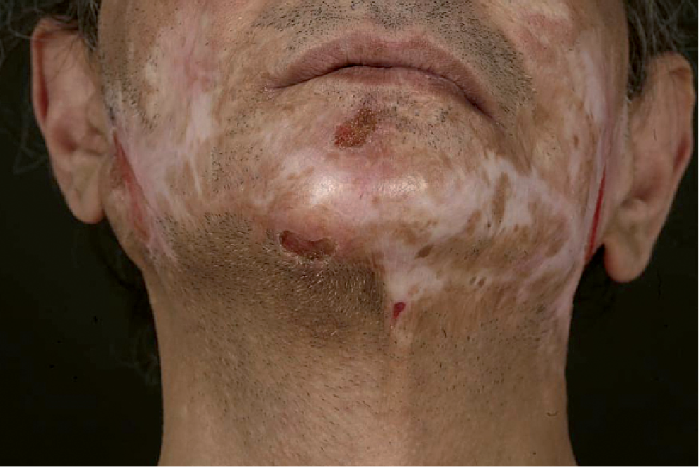
A 56-year-old man presented to our service a 6-year history of chronic facial pain, ulceration and subsequent scarring; which was preceded by an episode of a condition diagnosed in primary care as shingles. The left and right cheeks, left post-auricular area and chin were affected (Fig. 1). These changes had previously been diagnosed as secondary to dermatitis artefacta or acne excoriée. In the past he had used antidepressant medications and interacted with a psychologist, but terminated these therapies due to lack of perceived response.
Fig. 1. Bilateral ulceration and scarring affecting both cheeks, left postauricular region and chin.
Differential diagnoses of facial ulceration including the following were considered: malignancy, infection, vasculitis, pyoderma gangrenosum and psychodermatological diseases: dermatitis artefacta (DA), TTS and acne excoriée.
Swabs from active ulcers grew normal skin flora. Bloods revealed a microcytic anaemia, with no evidence of gastrointestinal bleeding found. Additional biochemical tests for pruritus were normal (liver function, urea and electrolytes, thyroid function, folate and Treponema serology). Skin biopsy noted only scarring of the epidermis. Magnetic resonance cranio-facial imaging showed mild chronic inflammation of the facial sinuses. Neurophysiology revealed bilateral trigeminal nerve dysfunction on both trigeminal nerve somatosensory evoked potentials and electromyography.
Our patient has been treated with simple analgesia (paracetamol and codeine); an antihistamine: hydroxyzine; selective serotonin re-uptake inhibitors: escitalopram, fluoxetine, paroxetine, sertraline; a tricyclic antidepressant: amitriptyline; antiepileptic drugs: gabapentin, pregabalin, topiramate and antipsychotics: quetiapine, olanzapine and amisulpride. All of these therapies achieved limited success. After lengthy consultation with pain services our patient is currently maintained on aripiprazole (5 mg daily), morphine sulphate (40 mg twice daily (BD)), pregabalin (300 mg BD), duloxetine (60 mg BD), oxcarbazepine 150 mg BD and topical lidocaine patches.
TTS is a rare but important cause of facial ulceration and consists of a triad of ulceration, anaesthesia and paraesthesia. TTS results from injury to the trigeminal nerve, which may be central or peripheral, and the resulting intractable dysasthesia leads to self-mutilating behaviour resulting in ulcers (1). These chronic ulcerating lesions can typically be observed in the nasal ala and paranasal areas but may be seen to involve any of the ophthalmic (V1) maxillary (V2) or mandibular (V3) nerve distributions (1, 2). Ulceration is characteristically unilateral and may occur as single or multiple lesions (3, 4).
The diagnosis is often made clinically. Patients are likely to report a preceding condition accounting for insult to the trigeminal nerve; namely stroke, trigeminal neuralgia, herpes zoster, meningioma, acoustic neuroma, encephalitis, syphilis or surgical procedures affecting the nerve (1). Patients are commonly misdiagnosed with DA, but the intractable facial sensations described by the patient and characteristic distribution should suggest a diagnosis of TTS (5). Histology is often non-specific. Neurophysiological studies can be helpful in evaluating function of the trigeminal nerve. Often a multi-disciplinary team (dermatologists, neurologists, psychiatrist and occasionally surgeons) is required to successfully treat patients. Patient education may help patients reverse destructive scratching habits (6). The most commonly administered medications are olanzapine, gabapentin and carbamazepine (5, 7). Occlusion dressings may be used to promote healing and surgical grafts or flaps may be necessary in order to cover ulcerated areas of skin. Treatment remains challenging and a number of therapies, including electrical stimulation, negative pressure therapy and autologous epidermal cell transplant have been investigated with varying degrees of success (1, 8–10).
The literature to date describes TTS as a unilateral condition and, to the best of the author’s knowledge, this is the first report of bilateral disease. We believe this patient has a clinically new entity, ATTS, which we are seeing in an increasing number of patients at our tertiary psychodermatology clinic. In ATTS, patients present with bilateral disease, presumably due to bilateral nerve insult, and this is confirmed by nerve conduction studies. Skin involvement is much more extensive than in TTS and may fluctuate in severity and location. Patients describe a sense of intense relief on skin picking, and they admit to voluntary picking (unlike in DA). Additionally, an obsessive-compulsive element often develops, which is much less a feature of TTS. ATTS is highly refractory to treatment, despite agents that are typically efficacious in TTS.
The authors declare no conflicts of interest.


 Click to show fullsize