


1Dermatology Department, 2Pneumology Department, 3Pathology Department, Saint-Louis Hospital, 1 avenue Claude Vellefaux, FR-75010 Paris, 4Paris VII Sorbonne Paris Cité University, Paris, and 5Pneumology Department, Compiègne Hospital, Compiègne, France. *E-mail: jean-david.bouaziz@aphp.fr
#These authors contributed equally and share senior authorship.
Accepted Apr 18, 2017; Epub ahead of print Apr 19, 2017
Eosinophilic granulomatosis with polyangiitis (Churg-Strauss) (EGPA) is an eosinophil-rich, necrotizing granulomatous inflammation, often involving the respiratory tract, and necrotizing vasculitis predominantly affecting small vessels and associated with asthma and eosinophilia (1). Allergic bronchopulmonary aspergillosis (ABPA) is characterized by asthma, type I hypersensitivity to Aspergillus, high total serum IgE serum level, eosinophilia, and bronchopulmonary involvement. We report here a case of a Koebner phenomenon revealing an EGPA in a patient who was recently diagnosed with ABPA.
A 54-year-old woman was followed in the department of pneumology for severe asthma. She was diagnosed with asthma and chronic sinusitis in 2006 and was treated with inhaled combined steroid therapy and long-acting bronchodilator. New explorations were conducted in 2015 because of recurrent asthma exacerbations. Chest computed tomography (CT) scan showed bronchial thickening, atelectasis and centrolobular micronodules. Sinus CT scan revealed bilateral maxillary and
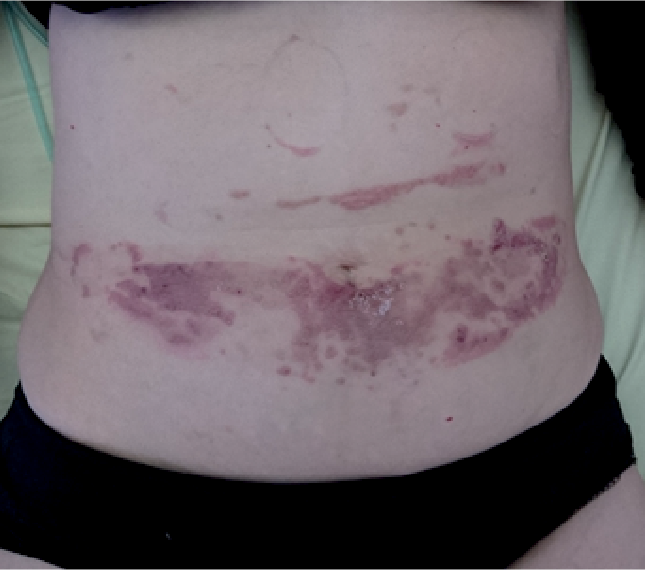
ethmoidal sinusitis. Laboratory tests showed an increased eosinophil cell count (2,500/mm3) (normal ranges (NR) < 500 cells/l), high total IgE level at 2,809 UI/ml (NR < 1,000 IU/ml), and positive Aspergillus-specific IgE level at 2.2kUi/l (NR< 0.35kUi/l). A diagnosis of ABPA was retained. Sinus bacteriological analysis was positive for Pseudomonas aeruginosa. An intravenous infusion of ceftazidime was started using a diffusing waist belt in association with oral ciprofloxacin. One week later, the patient developed non-pruritic purplish scaly papules around the waist strictly under the belt (Fig. 1).
Fig. 1. Purplish scaly papules round the waist.
She was admitted to the hospital as the dyspnoea worsened and because of new-onset epistaxis. Skin biopsy revealed palisading dermal extravascular necrotizing granulomas with eosinophils (Fig. 2). Nasal mid-turbinate histology also showed eosinophilic granulomas. Anti-neutrophil cytoplasmic antibodies (ANCA) were negative.
Fig. 2. Palisading dermal extravascular necrotizing granuloma. Hematoxylin-eosin staining; original magnification ×40.
The patient was thus diagnosed with EGPA with pulmonary, cutaneous and sinonasal involvement. The patients was treated with oral prednisone (1 mg/kg) with good efficiency (Fig. 3), which was well tolerated.
Fig. 3. Healing of skin lesions after oral corticotherapy.
The diagnosis of EGPA was established based on the following elements: association of asthma, high eosinophil cell count, extravascular necrotizing granuloma with eosinophils and cutaneous, pulmonary and sinonasal involvement. To date, only 9 cases associated with ABPA have been reported (2). Chronic exposure to fungal antigen and immune complex formation may play a role in the development of EGPA after ABPAs. No specific skin lesions during the course of ABPA have been described in the literature. EGPA cutaneous lesions are present in 40–80% of the patients, including purpura, nodules/papules or urticarial lesions and, less commonly, livedo reticularis, ulcerations and bullous lesions (3–5). Koebner phenomenon has been reported only once in an ANCA-positive patient, occurring on pyoderma gangrenosum scars (6). The Koebner pathogenesis is well known in psoriasis, but is poorly described in vasculitis (7). Nevertheless, skin lesions in granulomatous vasculitis are often located on the fingers and the elbows, which are frictional areas. The friction of the belt in our patient could have induced a local immune activation leading to skin lesions. We report here the first case of Koebner phenomenon in a patient with eosinophilic granulomatosis with polyangiitis.
The authors declare no conflicts of interest.


 Click to show fullsize
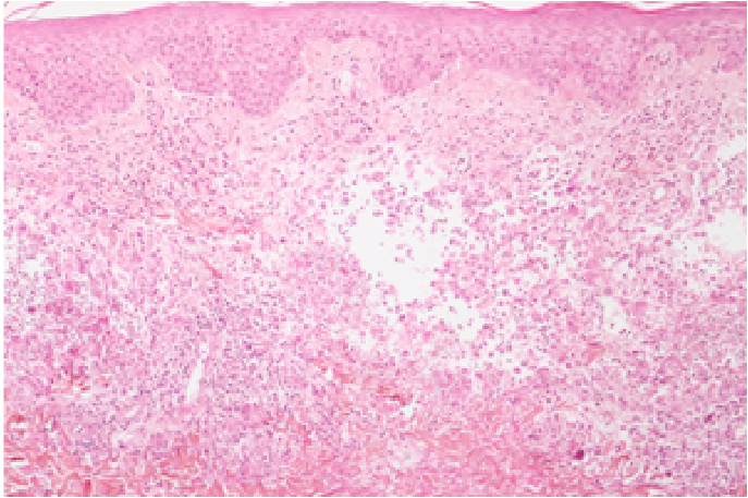 Click to show fullsize
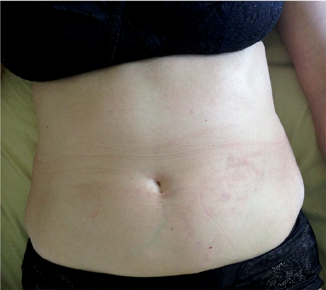 Click to show fullsize