


1Department of Cutaneous Medicine and Surgery, 2nd Floor South Tower, The Royal London Hospital and QMUL, Bart’s Health NHS Trust, Whitechapel, London E1 1BB, UK, 2CentroDerm® clinic, Wuppertal, and Faculty of Health, University Witten-Herdecke, Witten, Germany, 3Department of Dermato Cancerology, University of Nantes, Nantes, France, 4Department of Dermatology, Bellvitge Hospital, Barcelona, Spain, 5Manchester Academic Health Science Centre, MAHSC, Manchester University and Salford Royal NHS Foundation Trust, Royal Infirmary, The University of Manchester, Manchester, UK, 6Department of Dermatology, University of Modena and Reggio Emilia, Modena, 7Department of Dermatology, Catholic University of Rome, Rome, Italy, and 8Dermatology Unit, Virgen Macarena University Hospital, Seville, Spain. E-mail: r.cerio@qmul.ac.uk
Accepted Apr 27, 2017; Epub ahead of print Apr 27, 2017
*The Progressing Evidence in AK (PEAK) Working Group, formed to
identify and address existing educational needs in AK
Actinic keratosis (AK) is a chronic, progressive disease of the skin that has undergone long-term sun exposure. The affected areas contain visible and subclinical non-visible sun damage resulting in epidermal keratinocyte dysplasia, known by many as ‘field cancerisation’ (1), which is prone to AKs and sun-related skin cancer (2). Thus, visible AKs are clinical biomarkers for a photo-damaged field with subclinical damage associated with the unpredictable risk of progression to invasive squamous cell carcinoma (iSCC) (3). The aim of this multiexpert opinion article is to provide a discussion succinctly highlighting the clinical gaps for optimal management of AK: the lack of a universal definition and the need for a standardised grade assessment of AK/field cancerisation that also takes into account individual risk.
The prevalence of AK varies from 6–60%, depending on age, phototype and other predisposing risk factors (most notably immunosuppressed status, outdoor workers), and is increasing (1, 4), with a parallel increase in non-melanoma skin cancer (NMSC). AK presents a considerable socioeconomic burden, which will inevitably increase with an aging population (1, 4). To minimise this burden, AK should be recognised and treated, particularly in populations at high risk of NMSC. The goal of therapy should be to eliminate AK/field cancerisation (visible and non-visible subclinical lesions) to minimise risk of AK recurrence and potential progression to iSCC (5), although evidence for the latter is lacking. Some authors in specialised centres have shown the additional value of imaging methods in such management, particularly in visualising the evolution of subclinical lesions, which can be a challenge in current clinical practice (2).
Lesion-directed therapy (e.g. cryotherapy), treats only visible AKs, so field–directed treatment is necessary to treat subclinical damage, reduce AK recurrence rates and potentially minimise the risk of iSCC development (5). Recent guidelines recognise the importance of treating the entire field (5–7). However, cryotherapy alone remains the standard of care for treating AK patients with multiple lesions. This suggests that education, and growing evidence that treating the field is equally as important as treating visible AKs, will be instrumental in reducing the increasing disease burden. Shifting the treatment paradigm will require understanding the clinical gaps that need addressing.
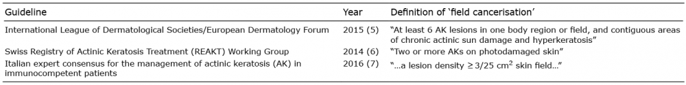
Firstly, there is a need for a standardised definition of AK field cancerisation in clinical, molecular and histopathological terms. Current guidelines define field cancerisation based on number of AK lesions and presence of surrounding photo-damaged skin (5–7). However, there are wide discrepancies within these criteria (Table I). Moreover, experts have voiced concerns over using a definition based on AK counts, as existing evidence suggests that any AK should be considered a marker of field change (8). A clearer and unambiguous definition of field cancerisation that is standardised and reproducible is required to support diagnosis and management, including treatment options and identification of ‘red flag’ signs of high-risk tumours. Physicians can only manage field cancerisation appropriately if they understand its characteristics and severity.
Table I. Various definitions of field cancerisation
A second clinical gap is the lack of a reproducible clinical global assessment scale for grading AK/field cancerisation. Current guidelines assess only the presence or absence of AK/field cancerisation, without a severity grading. A global assessment scale should include a clinical description of the key characteristics for each grade of severity to guide effectively identification, diagnosis and treatment decisions.
This clinical grading should be considered alongside modulating risk factors:
Through clinical grading based on disease severity and individual patient risk factors, a therapeutic algorithm that enables physicians to make informed treatment decisions would be valuable.
A relevant clinical challenge is the lack of direct evidence to date that AK can progress to iSCC, and that treating AK may prevent the risk of SCC. This is likely to impair the uptake of field therapy despite recommendations by current guidelines.
However, it seems reasonable to advocate field treat-ment in appropriate patients based on expert clinical judgement supported by:
In summary, addressing the lack of expert consensus on the definition and grading of AK/field cancerisation is crucial to aid physicians in their decision making and optimise appropriate management of AK.
Aiming to address such a need, the authors propose to undertake:
Editorial assistance from Lucid Group (funded by LEO Pharma) is gratefully acknowledged.
Conflict of interests: All authors act as consultants to Leo Pharma. GP reports receiving a research grant from Leo Pharma, and is a member of advisory boards for Leo Pharma, Roche and Galderma.


 Click to show fullsize