


Division of Dermatology, Department of Medicine of Sensory and Motor Organs, School of Medicine, Tottori University Faculty of Medicine, Yonago 683-8504, Japan. *E-mail: sugita@med.tottori-u.ac.jp
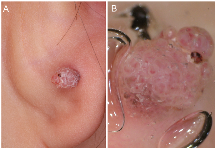
A 2-year-old boy was referred with a nodule on the back of his right auricle. The nodule had been present at birth and had enlarged gradually. Physical examination revealed a papillomatous, small, hypopigmented nodule 4×6 mm in diameter and 5 mm high (Fig. 1A). Dermoscopic examination revealed milky-red globules in a cobblestone pattern, a ring-like, whitish, scaly area, and dotted glomerular vessels (Fig. 1B). The patient had no identical or similar nodules on other sites.
What is your diagnosis? See next page for answer.
Fig. 1. (A) Hyperkeratotic, small, pink-to-red nodule, 4×6 mm in diameter and 5 mm high. (B) Dermoscopic findings. The nodule consisted of milky-red globules in a cobblestone pattern, a ring-like whitish scaly area, and dotted, glomerular vessels.
Acta Derm Venereol
Diagnosis: Congenital Spitz naevus
The nodule showed papillomatous and exophytic growth with surface hyperkeratosis. The constituent cells, with an epithelioid or spindle appearance, had proliferated in the dermis, exhibiting dilated vessels (Fig. 2A). These cells showed maturation at the bottom of the lesion with a few mitotic figures. Kamino bodies, which are commonly observed in Spitz naevus, were found (Fig. 2B). Tumour cells were positive for S-100 protein, melan-A and HMB-45. Based on these findings, a diagnosis was made of congenital Spitz naevus showing a hypopigmented verruciform appearance. The patient had no recurrence during a 6-month follow-up after excision.
Spitz naevus is a benign melanocytic tumour that occurs on the lower extremities and face at a relatively young age (1). To our knowledge, only 3 other articles have earlier described congenital Spitz naevus (2–4). Differential diagnosis includes pyogenic granuloma, verruca vulgaris, juvenile xanthogranuloma and, most importantly, amelanotic melanoma (5). Although verruca vulgaris in children usually presents as one or more papillomatous papules on the hands, feet or extremities, at the first visit we suspected that our patient had a verruca vulgaris.
Typical dermoscopic features of verruca vulgaris are densely multiple and packed papilla containing central, red or black, dotted or looped, vessels, surrounded by a whitish halo (6). In our patient, a ring-like whitish scaly area and dotted and glomerular vessels were seen. However, 22% of Spitz naevus cases also show dotted or looped vessels, as in verruca vulgaris (7). Thus, it is sometimes difficult to differentiate verruca vulgaris from Spitz naevus by dermoscopy, and further histopathological analysis is needed to make an accurate diagnosis.
Fig. 2. (A) Low-power magnification of a histopathological specimen shows exophytic growth with hyperkeratosis and papillomatosis (haematoxylin and eosin (HE) staining, ×40). (B) A high-power view shows proliferation of epithelioid and spindle cells in the dermis and Kamino bodies (yellow arrow) (HE staining, ×400).


 Click to show fullsize
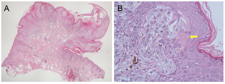 Click to show fullsize