


1Shandong Provincial Institute of Dermatology and Venereology, and 2Shandong Provincial Hospital for Skin Diseases, Shandong Academy of Medical Sciences, Jinan, Shandong, China. *E-mail: zhangfuren@hotmail.com
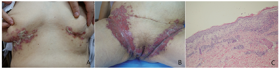
A 69-year-old woman presented with a 2-month history of painful lesions, which had initially appeared on the axillae, and had spread rapidly to the infra-mammary folds and groin. On physical examination there were erythematous papules coalescing into plaques and erosions in the intertriginous areas, oozing purulent fluid, including in the inframammary folds and groin (Fig. 1 A, B). There was no lymphadenopathy, lymphangitis or mucous membrane involvement. The patient had no other systemic symptoms. Results of laboratory tests, including urea, electrolytes, liver function and blood cell counts were normal. A skin biopsy was obtained, which showed the presence of sparse inflammatory infiltrates with numerous plasma cells (Fig. 1 C).
What is your diagnosis? See next page for answer.
Fig. 1. (A, B) Erythematous papules and erosions in the intertriginous areas, with massive amounts of purulent fluid in the inframammary fold and groin. (C) Skin biopsy showed the presence of a sparse number of plasma cells (original magnification, ×20).
As the histological findings of skin biopsy indicated a possible diagnosis of infectious disease, a specific immunohistochemical staining using antibodies against Treponema pallidum (TP) was performed and was strongly positive (Fig. 2 A, B). To support the diagnosis of syphilis, we subsequently ordered serological tests for syphilis, which showed a positive TRUST with a titre of 1:256 and positive TPPA. High load of TP in the lesion fluid was also identified by qPCR (Fig. 2C). A HIV test was negative. The patient was treated with penicillin G benzathine once a week for 3 weeks with dramatic improvement. She reported having had unprotected sexual contact only with her husband, who finally acknowledged that he had recently been diagnosed with syphilis and had been treated.
Fig. 2. (A, B) Immunohistochemistry stain of biopsy specimen (anti-Treponema pallidum (TP) polyclonal antibody immunostain (original magnification, ×20 and ×400). (C) High load of TP in the lesion fluid was identified by qPCR.
Due to the varied clinical manifestations, syphilis has been known as “the great imitator” and can mimic many skin disorders (1). The appearance of the eruption of secondary syphilis could be macular, maculopapular, nodular, nodule ulcerative or pustular (2), while the generalized lesions are usually associated with immunocompromised status. HIV can be associated with severe manifestation of secondary syphilis (3), such as malignant syphilis and erythema multiforme. The patient was HIV negative. Similar, but more localized, lesions have been reported as pustular syphilis, which is exceedingly rare and can present as miliary, acuminate, impetiginoid and rupioid forms (4).
For this patient, the clinical manifestation initially oriented to the diagnosis of Hailey-Hailey disease or Paget’s disease. A case with Hailey-Hailey disease presenting with very similar findings was reported recently (5). To the best of our knowledge, large erosions with purulent fluid on the intertriginous area have not been reported previously as symptoms for syphilis in the literature in English and Chinese. In this patient, the diagnosis of secondary syphilis was accomplished by positive serological test results, a histopathological pattern and positive immunohistochemical staining, and high load of TP from the lesion’s fluid tested by qPCR. Atypical cutaneous manifestation of syphilis may obscure the diagnosis and delay the provision of appropriate treatment. Clinicians must remain vigilant and consider syphilis in the differential diagnosis.


 Click to show fullsize
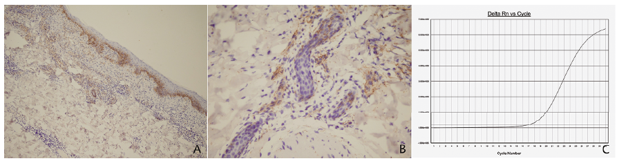 Click to show fullsize