


Department of Dermatology, Medical Center – University of Freiburg, DE-79104 Freiburg, Germany. E-mail: rudolf.happle@uniklinik-freiburg.de
Accepted Oct 15, 2018; Epub ahead of print Oct 15, 2018
In autosomal dominant disorders, 2 types of segmental mosaicism can be distinguished. The well-known type 1 results from a postzygotic mutation in an otherwise healthy embryo, whereas type 2 originates in a heterozygous embryo due to early loss of the corresponding wild-type allele, usually by somatic recombination (1). This mechanism gives rise to pronounced segmental involvement being superimposed on the non-segmental lesions of a given phenotype. Molecular proof of type 2 segmental mosaicism (T2SM) has been provided in many autosomal dominant skin disorders, including Hailey-Hailey disease, Darier’s disease, neurofibromatosis 1, Gorlin syndrome, and PTEN hamartoma syndrome (2).
Noonan syndrome (NS) is an autosomal dominant trait characterized by short stature, dysmorphic facial appearance, low-set, posteriorly rotated ears, and congenital heart defects including pulmonic stenosis (3). The disorder (OMIM #163950) is genetically heterogeneous (4). Approximately 50% of cases are caused by a heterozygous mutation in PTPN11. The remaining cases are due to mutations in SOS1, KSRS, NRAS, BRAF, RIT1, SOS2, or LZTR1 (4). The objective of this article is to review some reports that document cutaneous features suggesting T2SM in children with NS.
Lacombe et al. (5) described a newborn boy with respiratory distress and typical features of NS, including hypertelorism, ptosis, low-set posteriorly rotated ears, micrognathia, cleft palate, bilateral cryptorchidism, and redundant nuchal skin. X-ray examination revealed 13 ribs with a supernumerary vertebra. Echocardiography revealed an aortic supravalvular stenosis with cardiomyopathy. Moreover, a 6×8 cm large patch of “molluscoid” tissue, covered with folded skin of normal colour, was noted in the left frontoparietal area of the scalp. The boy died from cardiac failure at the age of 3 weeks. Histopathological examination of the unusual segmental scalp lesion showed hyperplasia of normal skin with proliferation of “radiating adipocytes”. The authors categorized this lesion as “nevoid cutis verticis gyrata”. The patient’s mother had typical features of NS. Later, the same case was published in French by Masson et al. (6). Histopathologically, they described “an organoid aspect with thickened subcutis of which the lobules are oriented in a radial pattern above the fascia.” The authors noted that the “nevoid cutis verticis gyrata” was clinically reminiscent of a large plexiform neurofibroma, although the histopathological findings excluded this diagnosis.
Fox et al. (7) documented a 2-day-old African-American girl who had mild respiratory distress, a III/VI holosystolic murmur at the left sternal border, hypertelorism, epicanthal folds, mild proptosis, a depressed nasal bridge, posteriorly rotated and low-set ears, a high-arched palate, redundant nuchal folds, and wide-spaced nipples. Moreover, she had a 6×6 cm “plaque of thick furrows, folds and relative alopecia in the left frontoparietal scalp.” The authors felt that this skin lesion was most consistent with “cutis verticis gyrata” in a patient with NS. The patient’s mother had non-segmental features of NS. The authors believed that “resolved intrauterine scalp lymphedema may account for the clinical presentation of cutis verticis gyrata in patients with Noonan and Turner syndrome.”
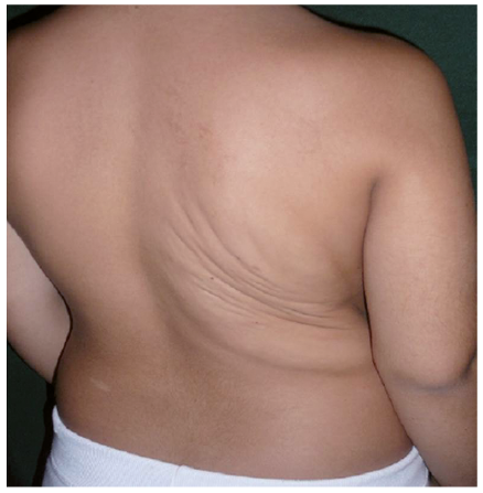
Boente et al. (8) reported a 10-year-old girl with typical features of NS, including triangular facial appearance, small low-set, posteriorly rotated ears, micrognathia, curly hair, low nuchal hairline, and webbed short neck. Moreover, she had in the right dorsal area a large plaque consisting of thick folds and deep furrows (Fig. 1). Microscopic examination of a biopsy obtained from this segmental lesion showed a normal epidermis “with disorganization of the dermal collagen”. The authors concluded that the unilateral “cutis gyrata” on the girl’s back “could also be considered a sign of the lymphatic alteration within this syndrome.”
Fig. 1. Segmentally arranged skin lesion consisting of thick folds with deep furrows in a 10-year-old girl with Noonan syndrome (8). (Reproduced with license under Creative Commons Attribution).
These 3 cases may best be explained at present as possible examples of T2SM in children with NS. The strict unilaterality of the skin lesions can be taken as an argument against the supposition that they simply represent sequelae of intrauterine lymphedema. Future molecular research may show whether the hypothesis of T2SM holds true.
In this context, it may be worth mentioning that the term “cutis verticis gyrata” has also been used for the large segmental cephalic hamartomas, as sometimes noted in tuberous sclerosis (9), which are probably a manifestation of T2SM in that disorder (2). Moreover, Masson et al. (6) noted a clinical resemblance of the patient’s scalp lesion with a plexiform neurofibroma (tumeur royale de neurofibromatose), a tumour that can today be taken as an example of T2SM in neurofibromatosis 1 (10).


 Click to show fullsize