


Departments of 1Dermatology and 2Pathology, CHRU Tours, FR-37044 Tours Cedex 9, France, 3Department of Dermatology, CHU Yalgado-Ouédraogo, Ouagadougou, Burkina Faso, and 4Inserm U1253, University of Tours, Tours, France. E-mail: r.safar@chu-tours.fr
Accepted Mar 13, 2019; E-published Mar 14, 2019
Bowen’s disease (BD), or squamous-cell carcinoma (SCC) in situ, frequently presents as a persistent red scaly patch that grows slowly over time and may appear as an ulcer or thickened scar. It most commonly occurs on areas or high exposure to sunlight, such as the face, neck, chest, arms and legs. The risk of invasive disease is 3–5% (1). BD as actinic keratosis occurs more often in older patients with comorbidities and is frequently located on body sites with poor wound healing (1). Surgical removal is usually recommended. In some widespread lesions, surgical removal and reconstruction may be challenging. Photodynamic therapy (PDT) may be an alternative to surgery (2). We report here a case of extensive BD treated successfully with daylight PDT.
An 88-year-old man presented to our dermatology department in March 2017 with an erosive lesion on the scalp, present for 6 months. He had a 10-year history of actinic keratosis treated with cryotherapy and topical 5-fluorouracil, as well as surgical removal of one basal cell carcinoma and one squamous cell carcinoma. The erosive lesion had started as a small erythematous lesion that gradually increased in size and became ulcerated with no other symptoms. Different types of dressings were tried, with no improvement.
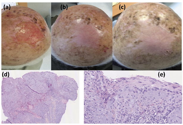
Clinical examination revealed an erosive plaque 10 cm in diameter, which was irregular with a sharply demarcated border (Fig. 1a). Dermoscopy revealed telangiectasia and glomeruloid vessels. A punched biopsy revealed an ulcerated lesion with fibrin necrotic leucocytes coating inflammatory granuloma associated with intense actinic damage, epidermal disorganization with dysplastic keratinocytes, individual dyskeratotic cells and increased mitotic activity with atypical mitotic figures (Fig. 1d, e). On the basis of clinical and histological examination, the lesion was diagnosed as BD.
Fig. 1. Pre- and post-treatment appearance of extensive Bowen’s disease on the scalp in an 88-year-old man. (a) Before daylight photodynamic therapy (PDT). (b) Complete healing at 3 months, and (c) 9 months after therapy. (d) Pre-treatment histology of the lesion, low-magnification periodic acid-Schiff staining, shows superficial ulceration and actinic damage (magnification ×50). (e) Higher-magnification (×400), haematoxylin phloxine saffron staining shows, in a small area of remaining epidermis, dyskeratotic cells with marked nuclear atypia and mitotic activity with no dermal extension, consistent with Bowen’s disease.
Because of the advanced age of the patient and the wide area and location of the lesion, alternatives to surgical removal were considered, and these corresponded with the patient’s preference. The patient refused conventional PDT because of the possible associated pain. Daylight PDT was performed in August 2017 at 14.00 h. First, sunscreen (SPF 50+) was applied on the patient’s scalp and face. Methyl aminolevulinate was then applied on the lesion. After 30 min, the patient stayed outdoors and exposed the treated area to sunlight for 2 h. After daylight exposure was completed, the patient was advised to wash off the residual cream and to stay at home for the rest of the day. Three months later, the patient had complete healing, with good cosmetic results (Fig. 1b), which persisted 9 months later (Fig. 1c). However, he still had multiple small lesions of actinic keratosis on the rest of the scalp, which were treated with another session of daylight PDT.
BD and actinic keratosis are both precursors of invasive SCC. Treatment of BD is based on the size and number of lesions, the general health of the patient, comorbidities and medication intake (3). Surgical excision allows for histological diagnosis and adequate control of the margins. Other options do not allow for histological control of the margins, including topical medication (5-fluorouracil, imiquimod, ingenol mebutate) or physical destruction using cryotherapy, curettage, irradiation, laser treatment and conventional PDT (4).
The European guidelines for conventional PDT include BD (4). Moreover, therapy guidelines recommend PDT as the treatment of choice for both large and small plaques of SCC in situ on poor healing sites and a good choice for large lesions on good healing sites (4, 5). However, these approaches are painful, particularly for large or numerous lesions. Daylight PDT, using daylight as the activating light source, is increasingly and effectively being used to treat actinic keratosis. Several trials have demonstrated that daylight PDT achieves similar response rates as conventional PDT in treating non-hyperkeratotic actinic keratosis (6, 7) in a nearly painless way (8). However, there are no recommendations for treating BD with daylight PDT (9).
Because the lesion reported here was large and totally erosive, it was hypothesized that daylight PDT could be the best treatment choice, combining efficacy and good tolerance. The patient was informed that daylight PDT is indicated to treat actinic keratosis, but is not a classical option in BD. The benefit/risk balance between surgery, radiation therapy, and conventional PDT, which are all indicated in BD, and daylight PDT, which is indicated in actinic keratosis, including the risks of treatment failure, local invasion and subsequent metastasis, were discussed.
To our knowledge, there are no reported cases of the use of PDT to treat extensive and ulcerated BD. In this patient, daylight PDT was effective for treating the BD, with good efficacy and high tolerability. Clinical follow-up was maintained because recurrence of the disease is still possible.
The authors have no conflicts of interest to declare.


 Click to show fullsize