


Departments of 1Dermatology and 3Pathology, CHR Orleans, FR-45000 Orléans, and 2Department of Dermatology, University of Tours, CHRU Tours, Tours, France. E-mail: marianela.ackerman@chr-orleans.fr
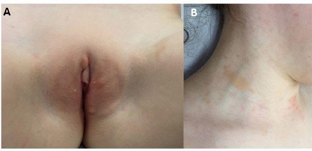
A 6-year-old girl with no significant medical history and who was not undergoing treatment, presented a 5-month history of multiple, scattered, asymptomatic, yellowish papulo-nodules with soft-elastic consistency in the vulva. Multiple, slightly papular, brownish plaques were on the inner side of her thighs and upper part of the back and neck (Fig. 1). Darier’s sign was negative for all lesions. Complete physical examination revealed no other anomaly, no other systemic complaints were present, and laboratory findings were normal.
A skin biopsy of a nodule was performed (Fig. 2).
What is your diagnosis? See next page for answer.
Fig. 1. (A) Nodules on the vulva and plaque on the thigh. (B) Plaques on the neck.
Fig. 2. (A) Haematoxylin-eosin staining 50×: deep and dense cellular infiltrate in dermis. (B) Giemsa staining 400×. (C) CD117 immunochemistry 25×.
Acta Derm Venereol 2019; XX: XX–XX.
Diagnosis: Vulvar involvement of skin mastocytosis
Microscopy examination revealed a dense interstitial infiltration of the deep dermis, mainly involving round cells with central nuclei and cytoplasm rich in metachromatic granules on Giemsa staining, with positive immunochemistry for CD117. Clinicopathological findings were consistent with a diagnosis of skin mastocytosis.
The vulvar location is exceptional; 17 cases were found in the literature, including some of vulvar mastocytoma and vulvar swelling, but other cases (1–8) featured this particular multiple papulo-nodular clinical aspect, which requires a differential diagnosis with Langerhans histiocytosis (9) and other causes of vulvar papulo-nodules, such as pseudo-verrucous papules linked to local irritation, condylomas, molluscum contagiosum, xanthomas and juvenile xanthogranulomas (5, 6).
Mastocytosis represents a group of disorders with an increased number of mast cells in tissues such as skin, bone marrow and gastrointestinal tract. The skin is the most frequently involved organ, with 3 main forms of cutaneous presentation in childhood (urticaria pigmentosa (maculopapular), nodular (mastocytoma) and diffuse (10, 11)). However, this classification is not always satisfactory, as in cases such as ours, that have different associated types of lesions.
Urticaria pigmentosa is the most frequent form in children; it may be suspected when skin examination reveals reddish-brown macules, papules, or small nodules (10, 11), as in our patient, which supported the clinical diagnosis. Darier’s sign is sometimes negative, as in the current case (11).
The histological findings are mast cell infiltrates in dermis, with metachromatic granules on Giemsa staining, and positive immunochemistry for CD117; this infiltrate could be superficial and sparse or deep and dense, as in our case, which could explain the different clinical lesions (7, 10).
Symptoms of mastocytosis in children are due to mast-cell degranulation and mediator release such as itch, palpitations, headache, episodic flushing, bronchospasm, diarrhoea or syncope, which can appear spontaneously or after mechanical, physical or chemical stimulation (4, 10). However, most children with skin mastocytosis remain asymptomatic or have only minimal degranulation symptoms (10).
The treatment aims to prevent or control the symptoms. Depending on the severity, the therapeutic options include follow-up and treatment with anti-histamines antiH1 and antiH2, and ketotifen as well as tyrosine kinase inhibitors, such as imatinib and dasatinib (10).
Our patient remained asymptomatic, therefore simple surveillance, and not mast cell degranulation stimulators, was indicated.
In conclusion, vulvar involvement is exceptional in skin mastocytosis, but it should be included as a possible diagnosis of vulvar nodules in children.


 Click to show fullsize
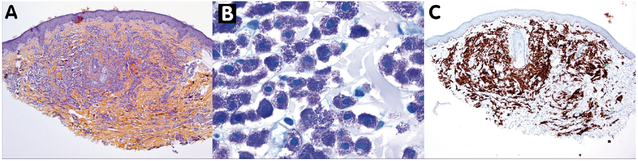 Click to show fullsize