


1Department of of Dermatology, Venereology and Allergology, and 2Department of Pathomorphology, Division of Pathomorphology, Wroclaw Medical University, 50-368 Wroclaw, Poland. E-mail: jacek.szepietowski@umed.wroc.pl
A 62-year-old woman, who worked as a seamstress, presented with an itchy, erythematous, scaly plaque on the dorsum of her fifth finger and the interdigital space 4/5 of her right, dominant hand. Physicians had diagnosed eczema 4 years previously, and had prescribed topical glucocorticosteroids. She reported having had breast cancer on the left side, which had been treated with mastectomy and adjuvant chemoradiotherapy 15 years previously, and had remained relapse-free.
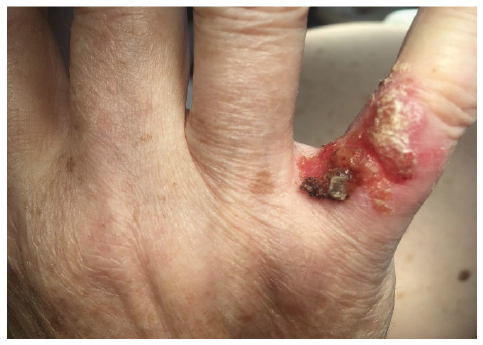
Differential diagnosis included skin cancer and chronic infection/inflammation. Biopsy revealed areas of dermal-subcutaneous necrobiosis surrounded by a granulomatous infiltrate exhibiting Langhans giant cells. PCR on paraffin-sections was positive for Mycobacterium tuberculosis. The initial tuberculostatic regimen consisted of rifampicin 10 mg/kg/day, isoniazid 5 mg/kg/day, pyrazinamide 25 mg/kg/day and ethambutol 15 mg/kg/day for a period of 2 months, to be continued with isoniazid and rifampicin for a further 4 months. The plaque improved with treatment, but did not resolve completely. At one year follow-up the patient presented with a progressive, ulcerated nodule at the same location (Fig. 1).
What is your diagnosis? See next page for answer.
Fig. 1. Erythemato-desquamative ulcerated lesion on the hand.
Acta Derm Venereol 2019; XX: XX–XX.
Diagnosis: Well-differentiated squamous cell carcinoma arising from lupus vulgaris, the most common form of cutaneous tuberculosis (synonymous: lupus carcinoma)
A skin biopsy was performed to exclude persistent tuberculosis or to search malignant transformation. Histopathological examination showed the features of a well-differentiated squamous cell carcinoma (SCC) (Fig. 2).
The patient underwent total excision with a security margin of 0.5 cm. On three-dimensional histopathology all surgical margins were free. The excisional defect was repaired with a full thickness graft from the left arm, with good functional and aesthetic result (Fig. 3). Baseline staging with axillary lymph node ultrasound and chest radiography did not detect any metastatic disease.
Lupus vulgaris (LV) is the most common form of cutaneous tuberculosis. The typical clinical appearance consists of a progressive plaque with elevated borders, central atrophy, and an apple jelly-like colour on pressure with a glass spatula (1). Incidence of malignant transformation has been reported to range from 0.5% to 10.5% (mean 4%) (2). “Lupus carcinoma”, i.e. SCC on active LV or LV scars typically occur decades after the first manifestation of LV. The elapsed time has been reported to range from 3 to 50 years (mean 28 years, median 35 years) (3, 4). Sunlight-exposed sites are predominant, suggesting cumulative toxicity of perpetuated repair mechanisms, immunosuppression and UV (1, 2, 5). In our patient the use of topical glucocorticosteroids for erroneously diagnosed “eczema” may have contributed to malignant transformation.
Today’s clinicians are currently not as familiar with cutaneous tuberculosis and LV as they were in the 19th and early 20th centuries. Nevertheless, LV and “lupus carcinoma” should be included in the differential diagnosis of chronic ulcerated, non-healing plaques on sun-exposed skin areas.
Fig. 2. Histological features of squamous cell carcinoma (haematoxylin and eosin; H&E ×100).
Fig. 3. Patient at follow-up 3 months after surgical procedure.


 Click to show fullsize
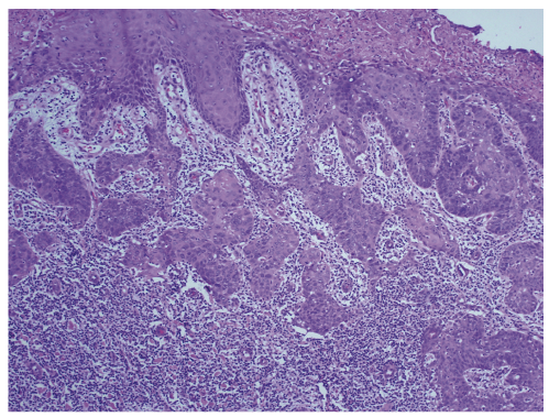 Click to show fullsize
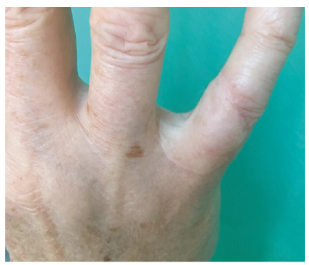 Click to show fullsize